 中文
中文 别名:Serine/threonine-protein kinase B-raf, Proto-oncogene B-Raf, p94, v-Raf murine sarcoma viral oncogene homolog B1, BRAF, BRAF1, RAFB1应用:WB,ICC
反应种属:Human
规格:50μl/100μl
| Description |
|---|
| This gene encodes a protein belonging to the raf/mil family of serine/threonine protein kinases. This protein plays a role in regulating the MAP kinase/ERKs signaling pathway, which affects cell division, differentiation, and secretion. Mutations in this gene are associated with cardiofaciocutaneous syndrome, a disease characterized by heart defects, mental retardation and a distinctive facial appearance. Mutations in this gene have also been associated with various cancers, including non-Hodgkin lymphoma, colorectal cancer, malignant melanoma, thyroid carcinoma, non-small cell lung carcinoma, and adenocarcinoma of lung. A pseudogene, which is located on chromosome X, has been identified for this gene. |
| Specification | |
|---|---|
| Aliases | Serine/threonine-protein kinase B-raf, Proto-oncogene B-Raf, p94, v-Raf murine sarcoma viral oncogene homolog B1, BRAF, BRAF1, RAFB1 |
| Entrez GeneID | 673 |
| Swissprot | P15056 |
| WB Predicted band size | 84.4kDa |
| Host/Isotype | Mouse IgG1 |
| Storage | Store at 4°C short term. Aliquot and store at -20°C long term. Avoid freeze/thaw cycles. |
| Species Reactivity | Human |
| Immunogen | This BRAF Monoclonal antibody is generated from mouse immunized with BRAF recombinant protein. |
| Formulation | Purified monoclonal antibody supplied in PBS with 0.05% sodium azide. This antibody is purified through a protein G column, eluted with high and low pH buffers and neutralized immediately, followed by dialysis against PBS. |
| Application | |
|---|---|
| WB | 1/1000 |
| ICC | 1/10-1/50 |
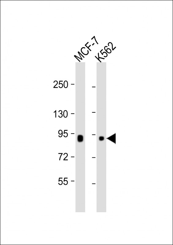 |
All lanes : Anti-BRAF Antibody at 1:1000 dilution Lane 1: MCF-7 whole cell lysate Lane 2: K562 whole cell lysate Lysates/proteins at 20 µg per lane. Secondary Predicted band size : 84 kDa Blocking/Dilution buffer: 5% NFDM/TBST. |
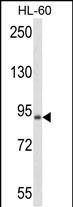 |
Western blot analysis of anti-BRAF Antibody in HL-60 cell line lysates (35μg/lane). BRAF (arrow) was detected using the purified Mab. |
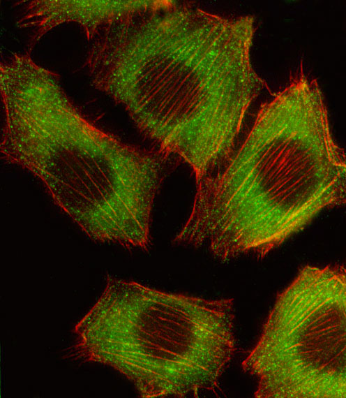 |
Fluorescent image of C2C12 cell stained with BRAF Antibody(Cat#P34057/SG100521B).C2C12 cells were fixed with 4% PFA (20 min), permeabilized with Triton X-100 (0.1%, 10 min), then incubated with BRAF primary antibody (1:25, 1 h at 37℃. For secondary antibody, Alexa Fluor® 488 conjugated donkey anti-mouse antibody (green) was used (1:400, 50 min at 37℃.Cytoplasmic actin was counterstained with Alexa Fluor® 555 (red) conjugated Phalloidin (7units/ml, 1 h at 37℃.BRAF immunoreactivity is localized to Cytoplasm significantly. |
本公司的所有产品仅用于科学研究或者工业应用等非医疗目的,不可用于人类或动物的临床诊断或治疗,非药用,非食用。
暂无评论
本公司的所有产品仅用于科学研究或者工业应用等非医疗目的,不可用于人类或动物的临床诊断或治疗,非药用,非食用。


发表回复