 中文
中文 别名:Density-regulated protein, DRP, Protein DRP1, Smooth muscle cell-associated protein 3, SMAP-3, DENR, DRP1应用:WB,IHC,ICC,FCM
反应种属:Human, Mouse
规格:50μl/100μl
| Description |
|---|
| May be involved in the translation of target mRNAs by scanning and recognition of the initiation codon. Involved in translation initiation; promotes recruitmnet of aminoacetyled initiator tRNA to P site of 40S ribosomes. Can promote release of deacylated tRNA and mRNA from recycled 40S subunits following ABCE1-mediated dissociation of post-termination ribosomal complexes into subunits. Plays a role in the modulation of the translational profile of a subset of cancer-related mRNAs when recruited to the translational initiation complex by the oncogene MCTS1. |
| Specification | |
|---|---|
| Aliases | Density-regulated protein, DRP, Protein DRP1, Smooth muscle cell-associated protein 3, SMAP-3, DENR, DRP1 |
| Entrez GeneID | 8562 |
| Swissprot | O43583 |
| WB Predicted band size | 22.1kDa |
| Host/Isotype | Mouse IgG2b |
| Storage | Store at 4°C short term. Aliquot and store at -20°C long term. Avoid freeze/thaw cycles. |
| Species Reactivity | Human, Mouse |
| Immunogen | This DENR antibody is generated from a mouse immunized with a recombinant protein of human DENR. |
| Application | |
|---|---|
| WB | 1/100-1/2000 |
| IHC | 1/100-1/500 |
| ICC | 1/25 |
| FCM | 1/25 |
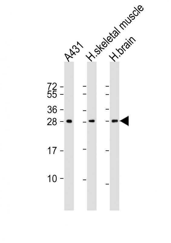 |
All lanes : Anti-DENR Antibody at 1:1000-1:2000 dilution Lane 1: A431 whole cell lysate Lane 2: human skeletal muscle lysate Lane 3: human brain lysate Lysates/proteins at 20 µg per lane. Secondary Predicted band size : 22 kDa Blocking/Dilution buffer: 5% NFDM/TBST. |
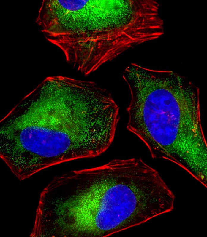 |
Immunofluorescent analysis of 4% paraformaldehyde-fixed, 0.1% Triton X-100 permeabilized HeLa (human cervical epithelial adenocarcinoma cell line) cells labeling DENR with P33379 at 1/25 dilution, followed by Dylight® 488-conjugated goat anti-mouse IgG secondary antibody at 1/200 dilution (green). Immunofluorescence image showing cytoplasm staining on HeLa cell line. Cytoplasmic actin is detected with Dylight® 554 Phalloidin at 1/100 dilution (red).The nuclear counter stain is DAPI (blue). |
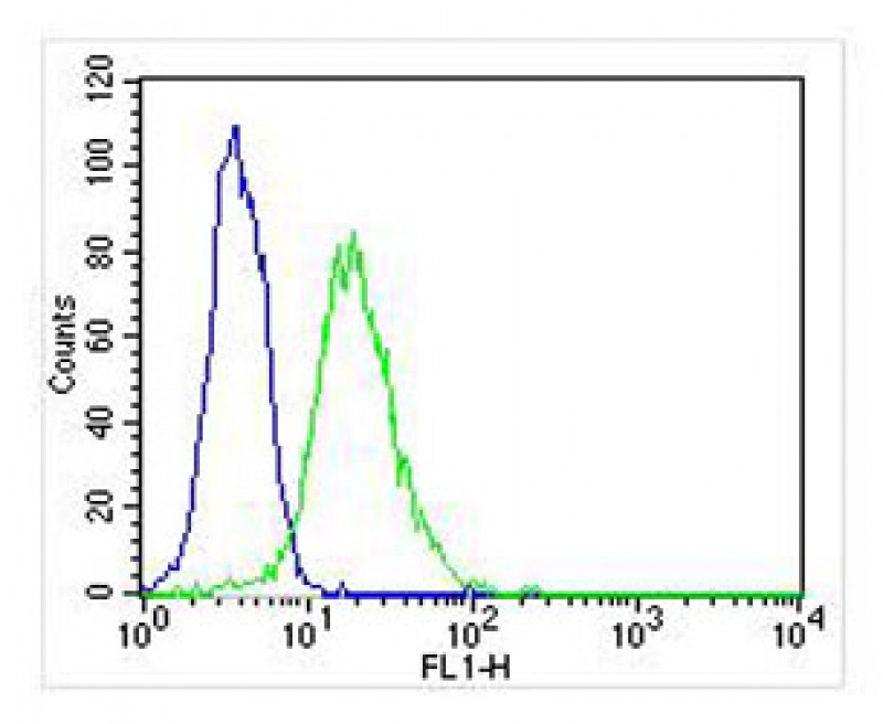 |
Overlay histogram showing Hela cells stained with P33379 (green line). The cells were fixed with 2% paraformaldehyde (10 min) and then permeabilized with 90% methanol for 10 min. The cells were then icubated in 2% bovine serum albumin to block non-specific protein-protein interactions followed by the antibody (P33379, 1:25 dilution) for 60 min at 37ºC. The secondary antibody used was Goat-Anti-Mouse IgG, DyLight® 488 Conjugated Highly Cross-Adsorbed(NA168821) at 1/400 dilution for 40 min at 37ºC. Isotype control antibody (blue line) was mouse IgG2b (1μg/1×10^6 cells) used under the same conditions. Acquisition of >10, 000 events was performed. |
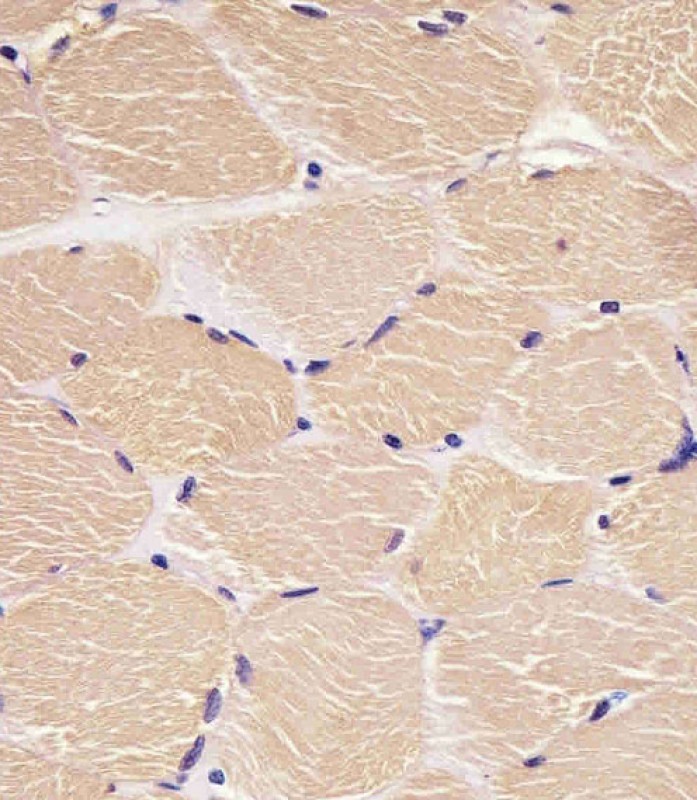 |
P33379 staining DENR in human skeletal muscle sections by Immunohistochemistry (IHC-P – paraformaldehyde-fixed, paraffin-embedded sections). Tissue was fixed with formaldehyde and blocked with 3% BSA for 0. 5 hour at room temperature; antigen retrieval was by heat mediation with a citrate buffer (pH6). Samples were incubated with primary antibody (1/25) for 1 hours at 37°C. A undiluted biotinylated goat polyvalent antibody was used as the secondary antibody. |
本公司的所有产品仅用于科学研究或者工业应用等非医疗目的,不可用于人类或动物的临床诊断或治疗,非药用,非食用。
暂无评论
本公司的所有产品仅用于科学研究或者工业应用等非医疗目的,不可用于人类或动物的临床诊断或治疗,非药用,非食用。


发表回复