 中文
中文 别名:Glial fibrillary acidic protein, GFAP, GFAP应用:WB,IHC,ICC
反应种属:Human, Mouse, Rat
规格:50μl/100μl
| Description |
|---|
| This gene encodes one of the major intermediate filament proteins of mature astrocytes. It is used as a marker to distinguish astrocytes from other glial cells during development. Mutations in this gene cause Alexander disease, a rare disorder of astrocytes in the central nervous system. Alternative splicing results in multiple transcript variants encoding distinct isoforms. |
| Specification | |
|---|---|
| Aliases | Glial fibrillary acidic protein, GFAP, GFAP |
| Entrez GeneID | 2670 |
| Swissprot | P14136 |
| WB Predicted band size | 49.9kDa |
| Host/Isotype | Mouse IgG2b |
| Storage | Store at 4°C short term. Aliquot and store at -20°C long term. Avoid freeze/thaw cycles. |
| Species Reactivity | Human, Mouse, Rat |
| Immunogen | This GFAP monoclonal antibody is generated from mouse immunized with GFAP recombinant protein. |
| Formulation | Purified monoclonal antibody supplied in PBS with 0.05% sodium azide. This antibody is purified through a protein G column, followed by dialysis against PBS. |
| Application | |
|---|---|
| WB | 1/4000 |
| IHC | 1/1000-1/2000 |
| ICC | 1/10-1/50 |
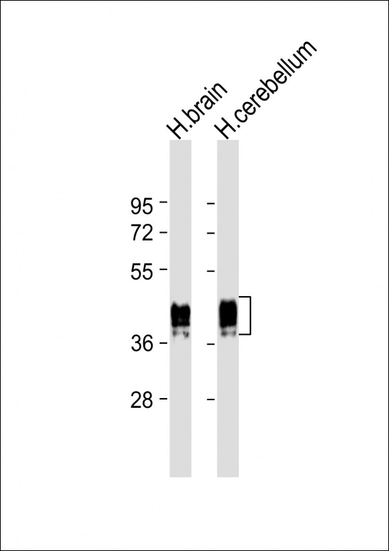 |
All lanes : Anti-GFAP Antibody at 1:4000 dilution Lane 1: human brain lysate Lane 2: human cerebellum lysate Lysates/proteins at 20 µg per lane. Secondary Predicted band size : 50 kDa Blocking/Dilution buffer: 5% NFDM/TBST. |
 |
Confocal immunofluorescent analysis of GFAP Antibody (Cat#P33582) with brain tissue followed by Alexa Fluor® 488-conjugated goat anti-mouse lgG (green). DAPI was used to stain the cell nuclear (blue). |
 |
Immunohistochemical analysis of paraffin-embedded human brain tissue using P33582 performed on the Leica® BOND RXm. Tissue was fixed with formaldehyde at room temperature; antigen retrieval was by heat mediation with a EDTA buffer (pH9. 0). Samples were incubated with primary antibody (1:1000) for 1 hours at room temperature. A undiluted biotinylated CRF Anti-Polyvalent HRP Polymer antibody was used as the secondary antibody. |
 |
Immunohistochemical analysis of paraffin-embedded Human brain section using Pink1(Cat#P33582). P33582 was diluted at 1:2000 dilution. A undiluted biotinylated goat polyvalent antibody was used as the secondary, followed by DAB staining. |
本公司的所有产品仅用于科学研究或者工业应用等非医疗目的,不可用于人类或动物的临床诊断或治疗,非药用,非食用。
暂无评论
本公司的所有产品仅用于科学研究或者工业应用等非医疗目的,不可用于人类或动物的临床诊断或治疗,非药用,非食用。


发表回复