 中文
中文 别名:Hypoxia-inducible factor 1-alpha inhibitor, 1.14.11.30, 1.14.11.n4, Factor inhibiting HIF-1, FIH-1, Hypoxia-inducible factor asparagine hydroxylase, HIF1AN, FIH1应用:WB,ICC
反应种属:Human
规格:50μl/100μl
| Description |
|---|
| Hydroxylates HIF-1 alpha at ‘Asp-803’ in the C-terminal transactivation domain (CAD). Functions as an oxygen sensor and, under normoxic conditions, the hydroxylation prevents interaction of HIF-1 with transcriptional coactivators including Cbp/p300- interacting transactivator. Involved in transcriptional repression through interaction with HIF1A, VHL and histone deacetylases. Hydroxylates specific Asn residues within ankyrin repeat domains (ARD) of NFKB1, NFKBIA, NOTCH1, ASB4, PPP1R12A and several other ARD-containing proteins. Also hydroxylates Asp and His residues within ARDs of ANK1 and TNKS2, respectively. Negatively regulates NOTCH1 activity, accelerating myogenic differentiation. Positively regulates ASB4 activity, promoting vascular differentiation. |
| Specification | |
|---|---|
| Aliases | Hypoxia-inducible factor 1-alpha inhibitor, 1.14.11.30, 1.14.11.n4, Factor inhibiting HIF-1, FIH-1, Hypoxia-inducible factor asparagine hydroxylase, HIF1AN, FIH1 |
| Entrez GeneID | 55662 |
| Swissprot | Q9NWT6 |
| WB Predicted band size | 40.3kDa |
| Host/Isotype | Mouse IgG1 |
| Storage | Store at 4°C short term. Aliquot and store at -20°C long term. Avoid freeze/thaw cycles. |
| Species Reactivity | Human |
| Immunogen | This HIF1AN antibody is generated from a mouse immunized with a KLH conjugated synthetic peptide between 1-349 amino acids from human HIF1AN. |
| Application | |
|---|---|
| WB | 1/2000 |
| ICC | 1/25 |
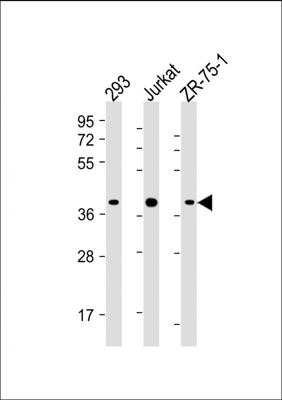 |
All lanes : Anti-HIF1AN Antibody (C-term) at 1:4000 dilution Lane 1: 293 whole cell lysate Lane 2: Jurkat whole cell lysate Lane 3: ZR-75-1 whole cell lysate Lysates/proteins at 20 µg per lane. Secondary Predicted band size : 40 kDa Blocking/Dilution buffer: 5% NFDM/TBST. |
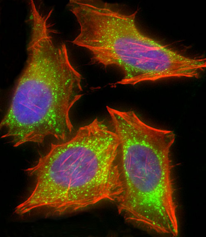 |
Immunofluorescent analysis of 4% paraformaldehyde-fixed, 0.1% Triton X-100 permeabilized HeLa (human cervical epithelial adenocarcinoma cell line) cells labeling HIF1AN with P34411 at 1/25 dilution, followed by Dylight® 488-conjugated goat anti-mouse IgG secondary antibody at 1/200 dilution (green). Immunofluorescence image showing mitochondrion staining on HeLa cell line. Cytoplasmic actin is detected with Dylight® 554 Phalloidin at 1/100 dilution (red). The nuclear counter stain is DAPI (blue). |
本公司的所有产品仅用于科学研究或者工业应用等非医疗目的,不可用于人类或动物的临床诊断或治疗,非药用,非食用。
暂无评论
本公司的所有产品仅用于科学研究或者工业应用等非医疗目的,不可用于人类或动物的临床诊断或治疗,非药用,非食用。


发表回复