 中文
中文 别名:KN motif and ankyrin repeat domain-containing protein 1, Ankyrin repeat domain-containing protein 15, Kidney ankyrin repeat-containing protein, KANK1, ANKRD15, KANK, KIAA0172应用:WB,IHC
反应种属:Human
规格:50μl/100μl
| Description |
|---|
| Involved in the control of cytoskeleton formation by regulating actin polymerization. Inhibits actin fiber formation and cell migration. Inhibits RhoA activity; the function involves phosphorylation through PI3K/Akt signaling and may depend on the competetive interaction with 14-3-3 adapter proteins to sequester them from active complexes. Inhibits the formation of lamellipodia but not of filopodia; the function may depend on the competetive interaction with BAIAP2 to block its association with activated RAC1. Inhibits fibronectin-mediated cell spreading; the function is partially mediated by BAIAP2. Inhibits neurite outgrowth. Involved in the establishment and persistence of cell polarity during directed cell movement in wound healing. In the nucleus, is involved in beta-catenin-dependent activation of transcription. Potential tumor suppressor for renal cell carcinoma. |
| Specification | |
|---|---|
| Aliases | KN motif and ankyrin repeat domain-containing protein 1, Ankyrin repeat domain-containing protein 15, Kidney ankyrin repeat-containing protein, KANK1, ANKRD15, KANK, KIAA0172 |
| Entrez GeneID | 23189 |
| Swissprot | Q14678 |
| WB Predicted band size | 147.3kDa |
| Host/Isotype | Mouse IgG1 |
| Storage | Store at 4°C short term. Aliquot and store at -20°C long term. Avoid freeze/thaw cycles. |
| Species Reactivity | Human |
| Immunogen | This KANK1 antibody is generated from a mouse immunized with a recombinant protein of human KANK1. |
| Application | |
|---|---|
| WB | 1/2000 |
| IHC | 1/100-1/500 |
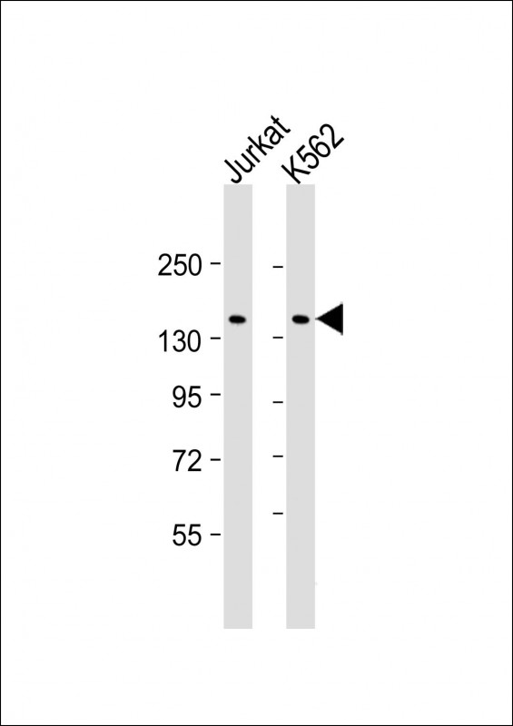 |
All lanes : Anti-KANK1 Antibody at 1:2000 dilution Lane 1: Jurkat whole cell lysate Lane 2: K562 whole cell lysate Lysates/proteins at 20 µg per lane. Secondary Predicted band size : 147 kDa Blocking/Dilution buffer: 5% NFDM/TBST. |
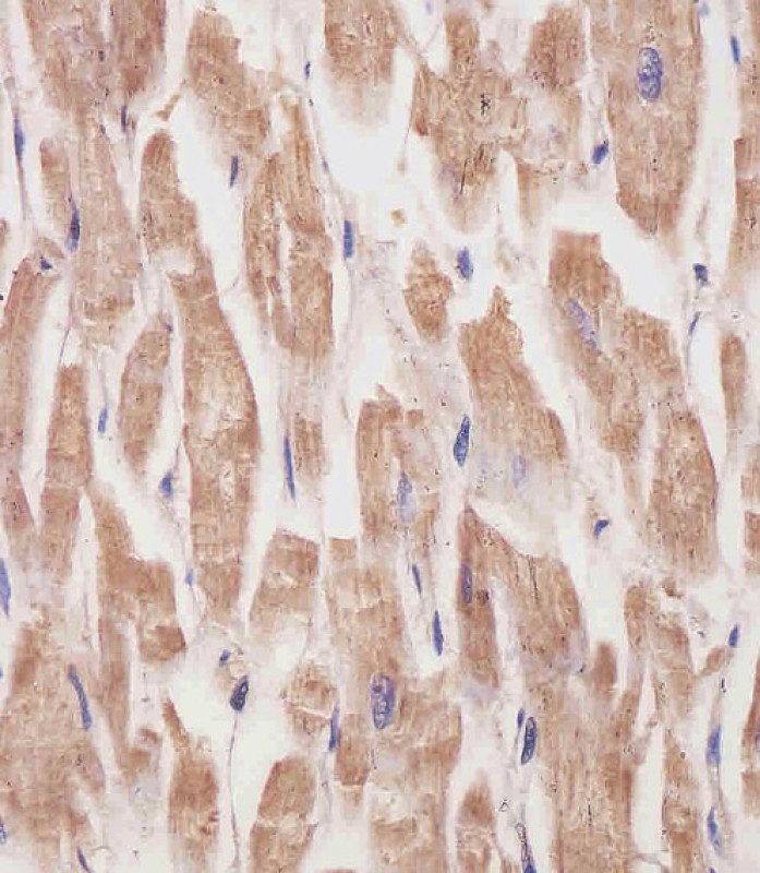 |
P34344 staining KANK1 in human heart tissue sections by Immunohistochemistry (IHC-P – paraformaldehyde-fixed, paraffin-embedded sections). Tissue was fixed with formaldehyde and blocked with 3% BSA for 0. 5 hour at room temperature; antigen retrieval was by heat mediation with a citrate buffer (pH6). Samples were incubated with primary antibody (1/25) for 1 hours at 37°C. A undiluted biotinylated goat polyvalent antibody was used as the secondary antibody. |
本公司的所有产品仅用于科学研究或者工业应用等非医疗目的,不可用于人类或动物的临床诊断或治疗,非药用,非食用。
暂无评论
本公司的所有产品仅用于科学研究或者工业应用等非医疗目的,不可用于人类或动物的临床诊断或治疗,非药用,非食用。


发表回复