 中文
中文 别名:Probable E3 ubiquitin-protein ligase makorin-2, 632-, RING finger protein 62, MKRN2, RNF62应用:WB,IHC,ICC,FCM
反应种属:Human, Mouse, Rat
规格:50μl/100μl
| Description |
|---|
| E3 ubiquitin ligase catalyzing the covalent attachment of ubiquitin moieties onto substrate proteins. |
| Specification | |
|---|---|
| Aliases | Probable E3 ubiquitin-protein ligase makorin-2, 632-, RING finger protein 62, MKRN2, RNF62 |
| Entrez GeneID | 23609 |
| Swissprot | Q9H000 |
| WB Predicted band size | 46.9kDa |
| Host/Isotype | Mouse IgG1 |
| Storage | Store at 4°C short term. Aliquot and store at -20°C long term. Avoid freeze/thaw cycles. |
| Species Reactivity | Human, Mouse, Rat |
| Immunogen | This MKRN2 antibody is generated from a mouse immunized with recombinant protein. |
| Application | |
|---|---|
| WB | 1/2000 |
| IHC | 1/100-1/500 |
| ICC | 1/25 |
| FCM | 1/25 |
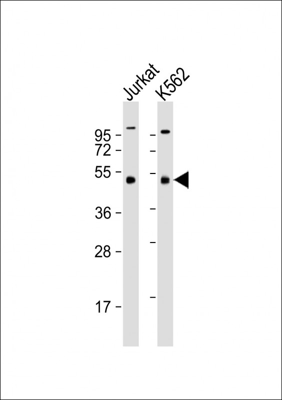 |
All lanes : Anti-MKRN2 Antibody at 1:4000 dilution Lane 1: Jurkat whole cell lysate Lane 2: K562 whole cell lysate Lysates/proteins at 20 µg per lane. Secondary Predicted band size : 47 kDa Blocking/Dilution buffer: 5% NFDM/TBST. |
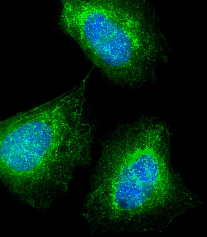 |
Immunofluorescent analysis of 4% paraformaldehyde-fixed, 0.1% Triton X-100 permeabilized U-2 OS (human osteosarcoma cell line) cells labeling MKRN2 with P33457 at 1/25 dilution, followed by Dylight® 488-conjugated goat anti-mouse IgG secondary antibody at 1/200 dilution (green). Immunofluorescence image showing cytoplasm and nucleus staining on U-2 OS cell line. The nuclear counter stain is DAPI (blue). |
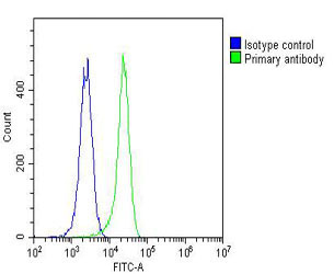 |
Overlay histogram showing K562 cells stained with P33457(green line). The cells were fixed with 2% paraformaldehyde (10 min) and then permeabilized with 90% methanol for 10 min. The cells were then icubated in 2% bovine serum albumin to block non-specific protein-protein interactions followed by the antibody (P33457, 1:25 dilution) for 60 min at 37ºC. The secondary antibody used was Goat-Anti-Mouse IgG, DyLight® 488 Conjugated Highly Cross-Adsorbed(OJ192088) at 1/200 dilution for 40 min at 37ºC. Isotype control antibody (blue line) was mouse IgG1 (1μg/1×10^6 cells) used under the same conditions. Acquisition of >10, 000 events was performed. |
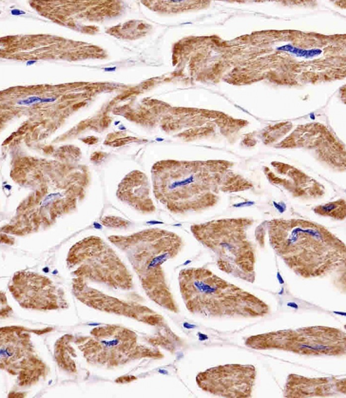 |
P33457 staining MKRN2 in human heart tissue sections by Immunohistochemistry (IHC-P – paraformaldehyde-fixed, paraffin-embedded sections). Tissue was fixed with formaldehyde and blocked with 3% BSA for 0. 5 hour at room temperature; antigen retrieval was by heat mediation with a citrate buffer (pH6). Samples were incubated with primary antibody (1/25) for 1 hours at 37°C. A undiluted biotinylated goat polyvalent antibody was used as the secondary antibody. |
本公司的所有产品仅用于科学研究或者工业应用等非医疗目的,不可用于人类或动物的临床诊断或治疗,非药用,非食用。
暂无评论
本公司的所有产品仅用于科学研究或者工业应用等非医疗目的,不可用于人类或动物的临床诊断或治疗,非药用,非食用。


发表回复