 中文
中文 别名:Phosphoglycerate kinase 1, 2.7.2.3, Cell migration-inducing gene 10 protein, Primer recognition protein 2, PRP 2, PGK1, PGKA应用:WB,IHC,FCM
反应种属:Human, Mouse, Rat
规格:50μl/100μl
| Description |
|---|
| In addition to its role as a glycolytic enzyme, it seems that PGK-1 acts as a polymerase alpha cofactor protein (primer recognition protein). |
| Specification | |
|---|---|
| Aliases | Phosphoglycerate kinase 1, 2.7.2.3, Cell migration-inducing gene 10 protein, Primer recognition protein 2, PRP 2, PGK1, PGKA |
| Entrez GeneID | 5230 |
| Swissprot | P00558 |
| WB Predicted band size | 44.6kDa |
| Host/Isotype | Mouse IgG2a |
| Storage | Store at 4°C short term. Aliquot and store at -20°C long term. Avoid freeze/thaw cycles. |
| Species Reactivity | Human, Mouse, Rat |
| Immunogen | This antibody is generated from a mouse immunized with a KLH conjugated synthetic peptide between 1-417 amino acids from human. |
| Application | |
|---|---|
| WB | 1/8000 |
| IHC | 1/100-1/500 |
| FCM | 1/25 |
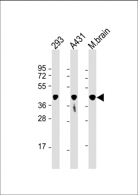 |
All lanes : Anti-PGK1 Antibody at 1:8000 dilution Lane 1: 293 whole cell lysate Lane 2: A431 whole cell lysate Lane 3: mouse brain lysate Lysates/proteins at 20 µg per lane. Secondary Predicted band size : 45 kDa Blocking/Dilution buffer: 5% NFDM/TBST. |
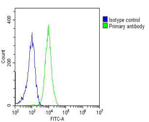 |
Overlay histogram showing Jurkat cells stained with P33969(green line). The cells were fixed with 2% paraformaldehyde (10 min) and then permeabilized with 90% methanol for 10 min. The cells were then icubated in 2% bovine serum albumin to block non-specific protein-protein interactions followed by the antibody (P33969, 1:25 dilution) for 60 min at 37ºC. The secondary antibody used was Goat-Anti-Mouse IgG, DyLight® 488 Conjugated Highly Cross-Adsorbed(OJ192088) at 1/200 dilution for 40 min at 37ºC. Isotype control antibody (blue line) was mouse IgG2a (1μg/1×10^6 cells) used under the same conditions. Acquisition of >10, 000 events was performed. |
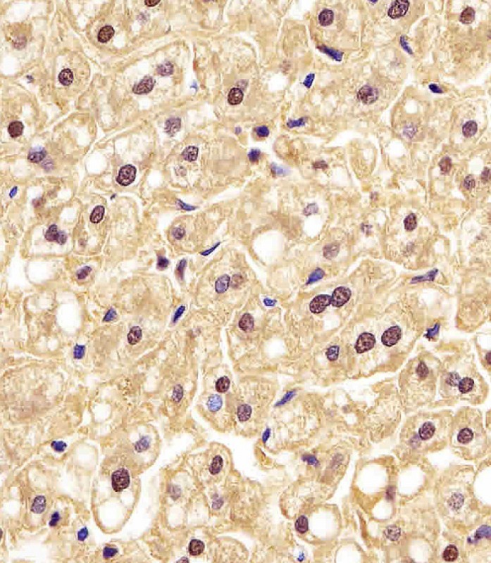 |
P33969 staining PGK1 in human liver tissue sections by Immunohistochemistry (IHC-P – paraformaldehyde-fixed, paraffin-embedded sections). Tissue was fixed with formaldehyde and blocked with 3% BSA for 0. 5 hour at room temperature; antigen retrieval was by heat mediation with a citrate buffer (pH6). Samples were incubated with primary antibody (1/25) for 1 hours at 37°C. A undiluted biotinylated goat polyvalent antibody was used as the secondary antibody. |
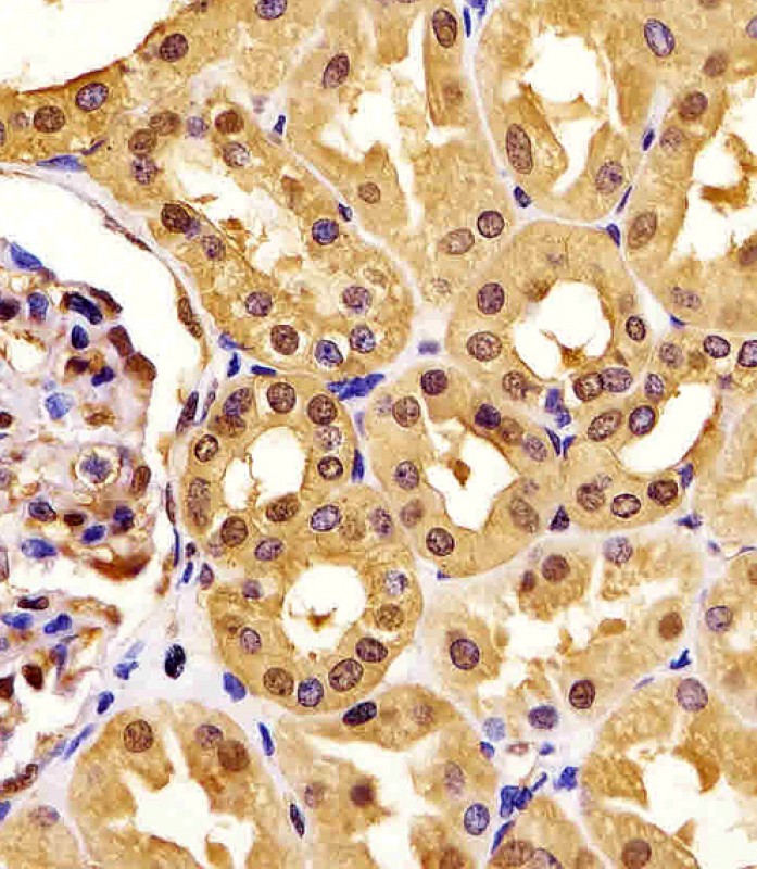 |
P33969 staining PGK1 in human kidney tissue sections by Immunohistochemistry (IHC-P – paraformaldehyde-fixed, paraffin-embedded sections). Tissue was fixed with formaldehyde and blocked with 3% BSA for 0. 5 hour at room temperature; antigen retrieval was by heat mediation with a citrate buffer (pH6). Samples were incubated with primary antibody (1/25) for 1 hours at 37°C. A undiluted biotinylated goat polyvalent antibody was used as the secondary antibody. |
本公司的所有产品仅用于科学研究或者工业应用等非医疗目的,不可用于人类或动物的临床诊断或治疗,非药用,非食用。
暂无评论
本公司的所有产品仅用于科学研究或者工业应用等非医疗目的,不可用于人类或动物的临床诊断或治疗,非药用,非食用。


发表回复