 中文
中文 别名:Proteasome subunit alpha type-5, Macropain zeta chain, Multicatalytic endopeptidase complex zeta chain, Proteasome zeta chain, PSMA5应用:WB,IHC,ICC
反应种属:Human, Mouse, Rat
规格:50μl/100μl
| Description |
|---|
| The proteasome is a multicatalytic proteinase complex with a highly ordered ring-shaped 20S core structure. The core structure is composed of 4 rings of 28 non-identical subunits; 2 rings are composed of 7 alpha subunits and 2 rings are composed of 7 beta subunits. Proteasomes are distributed throughout eukaryotic cells at a high concentration and cleave peptides in an ATP/ubiquitin-dependent process in a non-lysosomal pathway. An essential function of a modified proteasome, the immunoproteasome, is the processing of class I MHC peptides. This gene encodes a member of the peptidase T1A family, that is a 20S core alpha subunit. |
| Specification | |
|---|---|
| Aliases | Proteasome subunit alpha type-5, Macropain zeta chain, Multicatalytic endopeptidase complex zeta chain, Proteasome zeta chain, PSMA5 |
| Entrez GeneID | 5686 |
| Swissprot | P28066 |
| WB Predicted band size | 26.4kDa |
| Host/Isotype | Mouse IgG1 |
| Storage | Store at 4°C short term. Aliquot and store at -20°C long term. Avoid freeze/thaw cycles. |
| Species Reactivity | Human, Mouse, Rat |
| Immunogen | Purified His-tagged PSMA5 protein(Fragment) was used to produced this monoclonal antibody. |
| Formulation | Purified monoclonal antibody supplied in PBS with 0.05% sodium azide. This antibody is purified through a protein G column, followed by dialysis against PBS. |
| Application | |
|---|---|
| WB | 1/500-1/1000 |
| IHC | 1/100-1/500 |
| ICC | 1/25 |
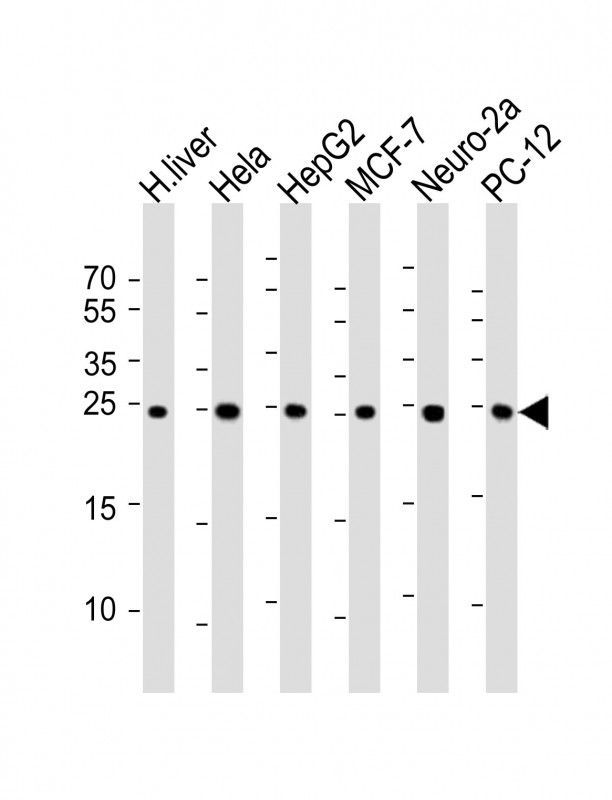 |
All lanes : Anti-PSMA5 Antibody at 1:1000 dilution Lane 1: human liver lysate Lane 2: Hela whole cell lysate Lane 3: HepG2 whole cell lysate Lane 4: MCF-7 whole cell lysate Lane 5: Neuro-2a whole cell lysate Lane 6: PC-12 whole cell lysate Lysates/proteins at 20 μg per lane. Secondary Predicted band size : 26 kDa. Blocking/Dilution buffer: 5% NFDM/TBST. |
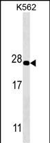 |
PSMA5 Antibody (Cat. #P33306) western blot analysis in K562 cell line lysates (35μg/lane).This demonstrates the PSMA5 antibody detected the PSMA5 protein (arrow). |
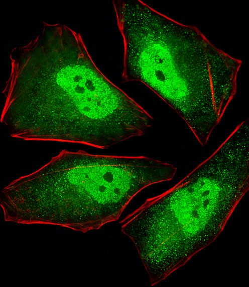 |
Fluorescent image of Hela cells stained with XAF1 PSMA5 Antibody(Cat#P33306). P33306 was diluted at 1:25 dilution. An Alexa Fluor® 488-conjugated goat anti-mouse lgG at 1:400 dilution was used as the secondary antibody (green). Cytoplasmic actin was counterstained with Alexa Fluor® 555 conjugated with Phalloidin (red). |
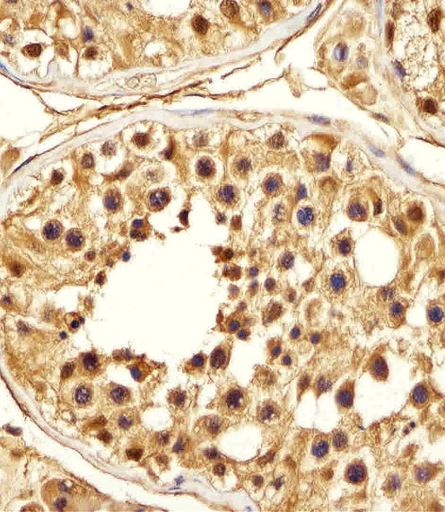 |
Immunohistochemical analysis of paraffin-embedded H. testis section using PSMA5 Antibody(Cat#P33306). P33306 was diluted at 1:25 dilution. A peroxidase-conjugated goat anti-mouse IgG at 1:400 dilution was used as the secondary antibody, followed by DAB staining. |
本公司的所有产品仅用于科学研究或者工业应用等非医疗目的,不可用于人类或动物的临床诊断或治疗,非药用,非食用。
暂无评论
本公司的所有产品仅用于科学研究或者工业应用等非医疗目的,不可用于人类或动物的临床诊断或治疗,非药用,非食用。


发表回复