 中文
中文 别名:Ras-related protein Rab-3A, RAB3A应用:WB,ICC,FCM
反应种属:Human, Mouse, Rat
规格:50μl/100μl
| Description |
|---|
| Involved in exocytosis by regulating a late step in synaptic vesicle fusion. Could play a role in neurotransmitter release by regulating membrane flow in the nerve terminal. |
| Specification | |
|---|---|
| Aliases | Ras-related protein Rab-3A, RAB3A |
| Entrez GeneID | 5864 |
| Swissprot | P20336 |
| WB Predicted band size | 30.0kDa |
| Host/Isotype | Mouse IgG1 |
| Storage | Store at 4°C short term. Aliquot and store at -20°C long term. Avoid freeze/thaw cycles. |
| Species Reactivity | Human, Mouse, Rat |
| Immunogen | This RAB3A antibody is generated from a mouse immunized with a recombinant protein. |
| Formulation | Purified monoclonal antibody supplied in PBS with 0.05% sodium azide. This antibody is purified through a protein G column, followed by dialysis against PBS. |
| Application | |
|---|---|
| WB | 1/500-1/2000 |
| ICC | 1/25 |
| FCM | 1/25 |
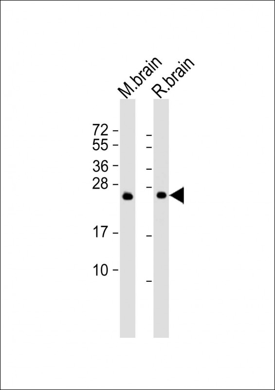 |
All lanes : Anti-RAB3A Antibody at 1:2000 dilution Lane 1: mouse brain lysate Lane 2: rat brain lysate Lysates/proteins at 20 μg per lane. Secondary Predicted band size : 25 kDa Blocking/Dilution buffer: 5% NFDM/TBST. |
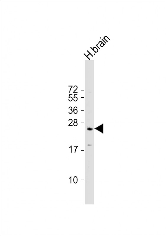 |
Anti-RAB3A Antibody at 1:500 dilution + human brain lysate
Lysates/proteins at 20 μg per lane. Secondary Predicted band size : 25 kDa Blocking/Dilution buffer: 5% NFDM/TBST. |
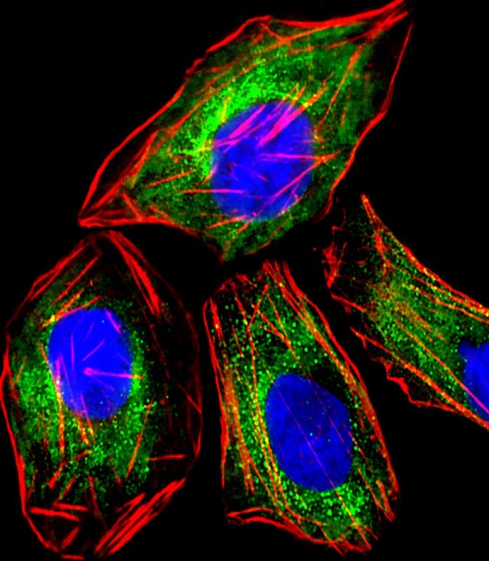 |
Immunofluorescent analysis of 4% paraformaldehyde-fixed, 0.1% Triton X-100 permeabilized PC-12 (rat adrenal phaeochromocytoma cell line) cells labeling RAB3A with P33311 at 1/25 dilution, followed by Dylight® 488-conjugated goat anti-mouse IgG secondary antibody at 1/200 dilution (green). Immunofluorescence image showing cytoplasm staining on PC-12 cell line. Cytoplasmic actin is detected with Dylight® 554 Phalloidin at 1/100 dilution (red).The nuclear counter stain is DAPI (blue). |
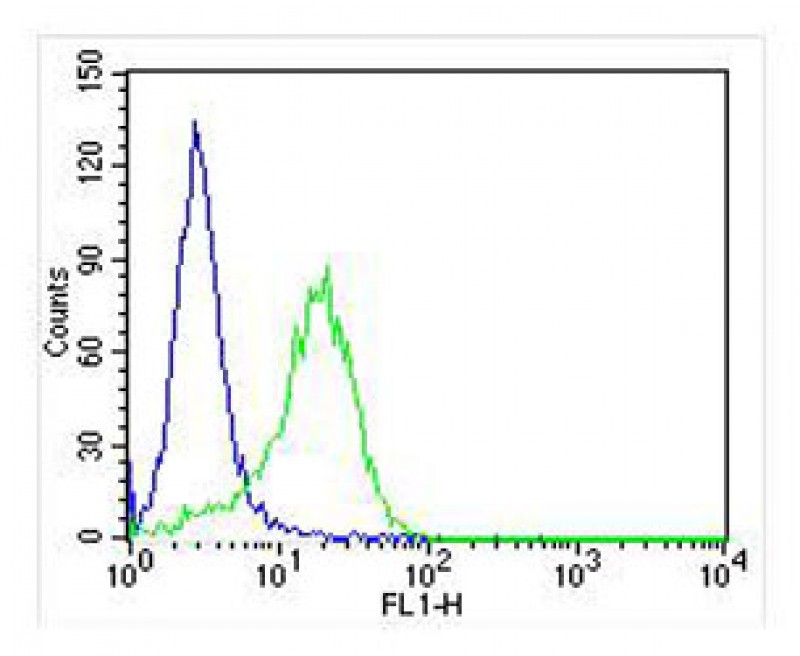 |
Overlay histogram showing PC-12 cells stained with P33311 (green line). The cells were fixed with 2% paraformaldehyde (10 min) and then permeabilized with 90% methanol for 10 min. The cells were then icubated in 2% bovine serum albumin to block non-specific protein-protein interactions followed by the antibody (P33311, 1:25 dilution) for 60 min at 37ºC. The secondary antibody used was Goat-Anti-Mouse IgG, DyLight® 488 Conjugated Highly Cross-Adsorbed(NA168821)) at 1/400 dilution for 40 min at 37ºC. Isotype control antibody (blue line) was mouse IgG (1μg/1×10^6 cells) used under the same conditions. Acquisition of >10, 000 events was performed. |
本公司的所有产品仅用于科学研究或者工业应用等非医疗目的,不可用于人类或动物的临床诊断或治疗,非药用,非食用。
暂无评论
本公司的所有产品仅用于科学研究或者工业应用等非医疗目的,不可用于人类或动物的临床诊断或治疗,非药用,非食用。


发表回复