 中文
中文 别名:Protein S100-A2, CAN19, Protein S-100L, S100 calcium-binding protein A2, S100A2, S100L应用:WB,IHC,ICC
反应种属:Human
规格:50μl/100μl
| Description |
|---|
| May function as calcium sensor and modulator, contributing to cellular calcium signaling. May function by interacting with other proteins, such as TPR-containing proteins, and indirectly play a role in many physiological processes. May also play a role in suppressing tumor cell growth. |
| Specification | |
|---|---|
| Aliases | Protein S100-A2, CAN19, Protein S-100L, S100 calcium-binding protein A2, S100A2, S100L |
| Entrez GeneID | 6273 |
| Swissprot | P29034 |
| WB Predicted band size | 11.1kDa |
| Host/Isotype | Mouse IgG1 |
| Storage | Store at 4°C short term. Aliquot and store at -20°C long term. Avoid freeze/thaw cycles. |
| Species Reactivity | Human |
| Immunogen | This S100A2 antibody is generated from a mouse immunized with a recombinant protein of human S100A2. |
| Application | |
|---|---|
| WB | 1/2000 |
| IHC | 1/100-1/500 |
| ICC | 1/25 |
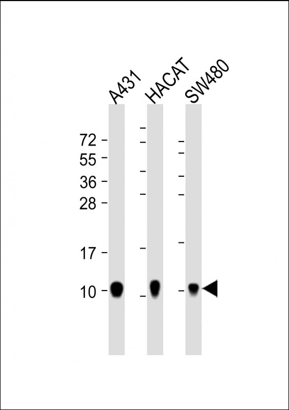 |
All lanes : Anti-S100A2 Antibody at 1:4000 dilution Lane 1: A431 whole cell lysate Lane 2: HACAT whole cell lysate Lane 3: SW480 whole cell lysate Lysates/proteins at 20 µg per lane. Secondary Predicted band size : 11 kDa Blocking/Dilution buffer: 5% NFDM/TBST. |
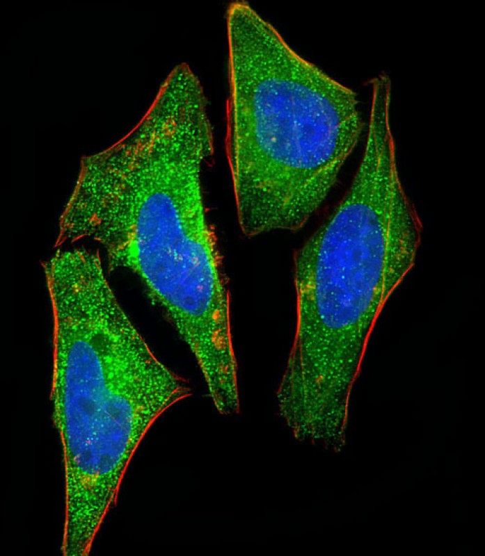 |
Immunofluorescent analysis of 4% paraformaldehyde-fixed, 0.1% Triton X-100 permeabilized HeLa (human cervical epithelial adenocarcinoma cell line) cells labeling S100A2 with P33515 at 1/25 dilution, followed by Dylight® 488-conjugated goat anti-mouse IgG secondary antibody at 1/200 dilution (green). Immunofluorescence image showing cytoplasm and nucleus staining on HeLa cell line. Cytoplasmic actin is detected with Dylight® 554 Phalloidin at 1/100 dilution (red).The nuclear counter stain is DAPI (blue). |
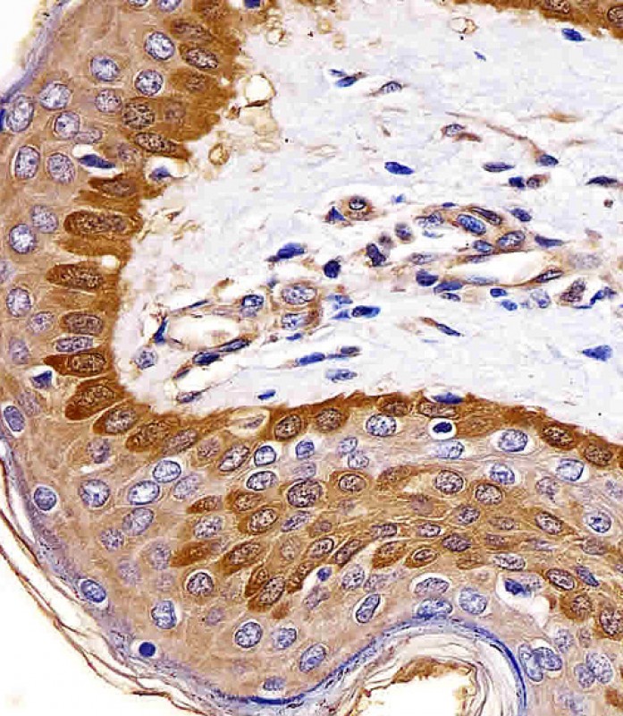 |
P33515 staining S100A2 in human skin tissue sections by Immunohistochemistry (IHC-P – paraformaldehyde-fixed, paraffin-embedded sections). Tissue was fixed with formaldehyde and blocked with 3% BSA for 0. 5 hour at room temperature; antigen retrieval was by heat mediation with a citrate buffer (pH6). Samples were incubated with primary antibody (1/25) for 1 hours at 37°C. A undiluted biotinylated goat polyvalent antibody was used as the secondary antibody. |
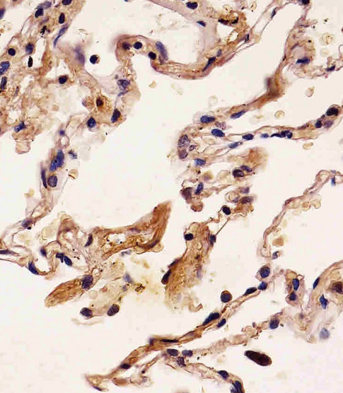 |
P33515 staining S100A2 in human lung tissue sections by Immunohistochemistry (IHC-P – paraformaldehyde-fixed, paraffin-embedded sections). Tissue was fixed with formaldehyde and blocked with 3% BSA for 0. 5 hour at room temperature; antigen retrieval was by heat mediation with a citrate buffer (pH6). Samples were incubated with primary antibody (1/25) for 1 hours at 37°C. A undiluted biotinylated goat polyvalent antibody was used as the secondary antibody. |
本公司的所有产品仅用于科学研究或者工业应用等非医疗目的,不可用于人类或动物的临床诊断或治疗,非药用,非食用。
暂无评论
本公司的所有产品仅用于科学研究或者工业应用等非医疗目的,不可用于人类或动物的临床诊断或治疗,非药用,非食用。


发表回复