 中文
中文 别名:Alpha-2-HS-glycoprotein, Alpha-2-Z-globulin, Ba-alpha-2-glycoprotein, Fetuin-A, Alpha-2-HS-glycoprotein chain A, Alpha-2-HS-glycoprotein chain B, AHSG, FETUA应用:WB,ICC,FCM
反应种属:Human
规格:50μl/100μl
| Description |
|---|
| Alpha2-HS glycoprotein (AHSG), a glycoprotein present in the serum, is synthesized by hepatocytes. The AHSG molecule consists of two polypeptide chains, which are both cleaved from a proprotein encoded from a single mRNA. It is involved in several functions, such as endocytosis, brain development and the formation of bone tissue. The protein is commonly present in the cortical plate of the immature cerebral cortex and bone marrow hemopoietic matrix, and it has therefore been postulated that it participates in the development of the tissues. However, its exact significance is still obscure. |
| Specification | |
|---|---|
| Aliases | Alpha-2-HS-glycoprotein, Alpha-2-Z-globulin, Ba-alpha-2-glycoprotein, Fetuin-A, Alpha-2-HS-glycoprotein chain A, Alpha-2-HS-glycoprotein chain B, AHSG, FETUA |
| Entrez GeneID | 197 |
| Swissprot | P02765 |
| WB Predicted band size | 39.4kDa |
| Host/Isotype | Rabbit IgG |
| Storage | Store at 4°C short term. Aliquot and store at -20°C long term. Avoid freeze/thaw cycles. |
| Species Reactivity | Human |
| Immunogen | This AHSG antibody is generated from rabbits immunized with a KLH conjugated synthetic peptide between 247-276 amino acids from the C-terminal region of human AHSG. |
| Formulation | Purified polyclonal antibody supplied in PBS with 0.05% sodium azide. This antibody is purified through a protein A column, followed by peptide affinity purification. |
| Application | |
|---|---|
| WB | 1/2000 |
| ICC | 1/25 |
| FCM | 1/25 |
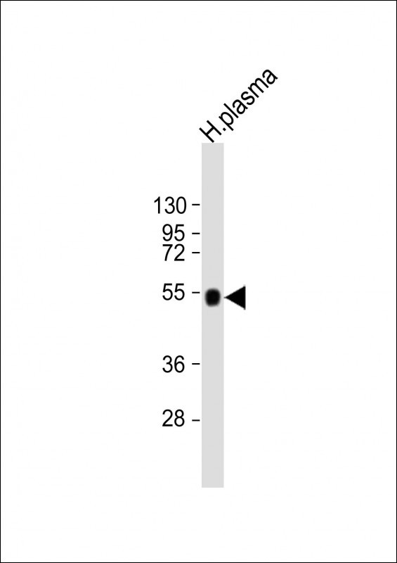 |
Anti-AHSG Antibody (C-term) at 1:2000 dilution + human plasma lysate
Lysates/proteins at 20 µg per lane. Secondary Predicted band size : 39 kDa Blocking/Dilution buffer: 5% NFDM/TBST. |
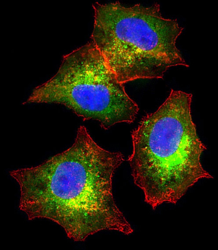 |
Immunofluorescent analysis of 4% paraformaldehyde-fixed, 0.1% Triton X-100 permeabilized HepG2 (human liver hepatocellular carcinoma cell line) cells labeling AHSG with P33383 at 1/25 dilution, followed by Dylight® 488-conjugated goat anti-rabbit IgG secondary antibody at 1/200 dilution (green). Immunofluorescence image showing cytoplasm staining on HepG2 cell line. Cytoplasmic actin is detected with Dylight® 554 Phalloidin at 1/100 dilution (red).The nuclear counter stain is DAPI (blue). |
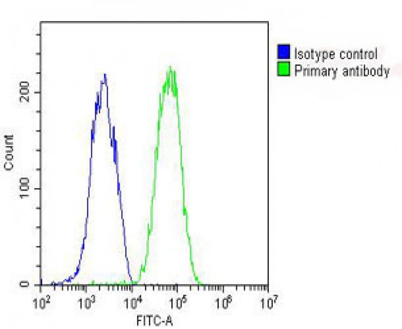 |
Overlay histogram showing HepG2 cells stained with P33383 (green line). The cells were fixed with 2% paraformaldehyde (10 min) and then permeabilized with 90% methanol for 10 min. The cells were then icubated in 2% bovine serum albumin to block non-specific protein-protein interactions followed by the antibody (P33383, 1:25 dilution) for 60 min at 37ºC. The secondary antibody used was Goat-Anti-Rabbit IgG, DyLight® 488 Conjugated Highly Cross-Adsorbed(OH191631) at 1/200 dilution for 40 min at 37ºC. Isotype control antibody (blue line) was rabbit IgG (1μg/1×10^6 cells) used under the same conditions. Acquisition of >10, 000 events was performed. |
本公司的所有产品仅用于科学研究或者工业应用等非医疗目的,不可用于人类或动物的临床诊断或治疗,非药用,非食用。
暂无评论
本公司的所有产品仅用于科学研究或者工业应用等非医疗目的,不可用于人类或动物的临床诊断或治疗,非药用,非食用。


发表回复