 中文
中文 别名:AP-1 complex subunit mu-1, AP-mu chain family member mu1A, Adaptor protein complex AP-1 subunit mu-1, Adaptor-related protein complex 1 subunit mu-1, Clathrin assembly protein complex 1 mu-1 medium chain 1, Clathrin coat assembly protein AP47, Clathrin coat-associated protein AP47, Golgi adaptor HA1/AP1 adaptin mu-1 subunit, Mu-adaptin 1, Mu1A-adaptin, AP1M1, CLTNM应用:WB,ICC,FCM
反应种属:Human, Mouse, Rat
规格:50μl/100μl
| Description |
|---|
| The protein encoded by this gene is the medium chain of the trans-Golgi network clathrin-associated protein complex AP-1. The other components of this complex are beta-prime-adaptin, gamma-adaptin, and the small chain AP1S1. This complex is located at the Golgi vesicle and links clathrin to receptors in coated vesicles. These vesicles are involved in endocytosis and Golgi processing. Alternatively spliced transcript variants encoding distinct protein isoforms have been found for this gene. [provided by RefSeq]. |
| Specification | |
|---|---|
| Aliases | AP-1 complex subunit mu-1, AP-mu chain family member mu1A, Adaptor protein complex AP-1 subunit mu-1, Adaptor-related protein complex 1 subunit mu-1, Clathrin assembly protein complex 1 mu-1 medium chain 1, Clathrin coat assembly protein AP47, Clathrin coat-associated protein AP47, Golgi adaptor HA1/AP1 adaptin mu-1 subunit, Mu-adaptin 1, Mu1A-adaptin, AP1M1, CLTNM |
| Entrez GeneID | 8907 |
| Swissprot | Q9BXS5 |
| WB Predicted band size | 48.6kDa |
| Host/Isotype | Rabbit IgG |
| Storage | Store at 4°C short term. Aliquot and store at -20°C long term. Avoid freeze/thaw cycles. |
| Species Reactivity | Human, Mouse, Rat |
| Immunogen | This AP1M1 antibody is generated from rabbits immunized with a KLH conjugated synthetic peptide between 205-234 amino acids from the Central region of human AP1M1. |
| Formulation | Purified polyclonal antibody supplied in PBS with 0.05% sodium azide. This antibody is purified through a protein A column, followed by peptide affinity purification. |
| Application | |
|---|---|
| WB | 1/1000 |
| ICC | 1/25 |
| FCM | 1/25 |
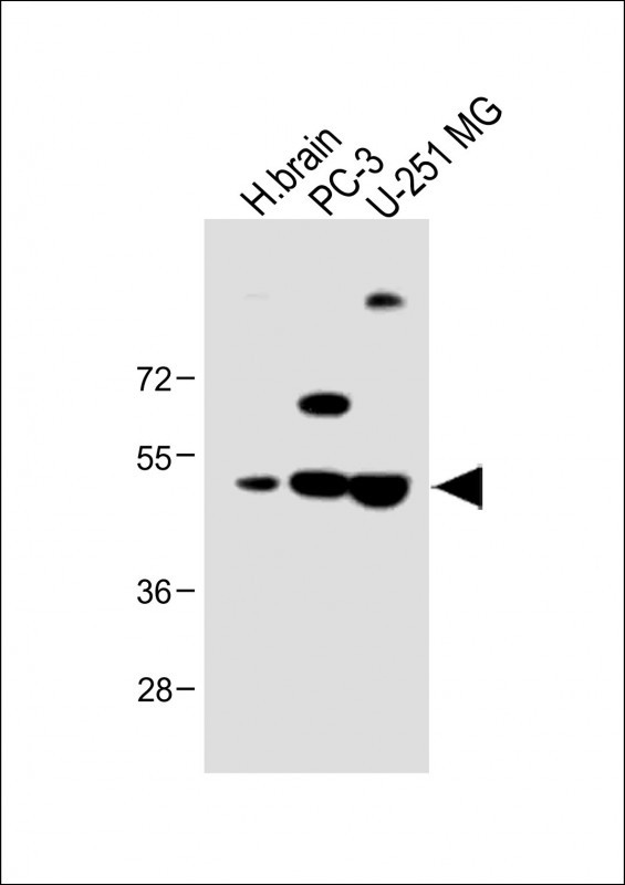 |
All lanes : Anti-AP1M1 Antibody (Center) at 1:1000 dilution Lane 1: Human brain lysate Lane 2: PC-3 whole cell lysate Lane 3: U-251 MG whole cell lysate Lysates/proteins at 20 µg per lane. Secondary Predicted band size : 49 kDa Blocking/Dilution buffer: 5% NFDM/TBST. |
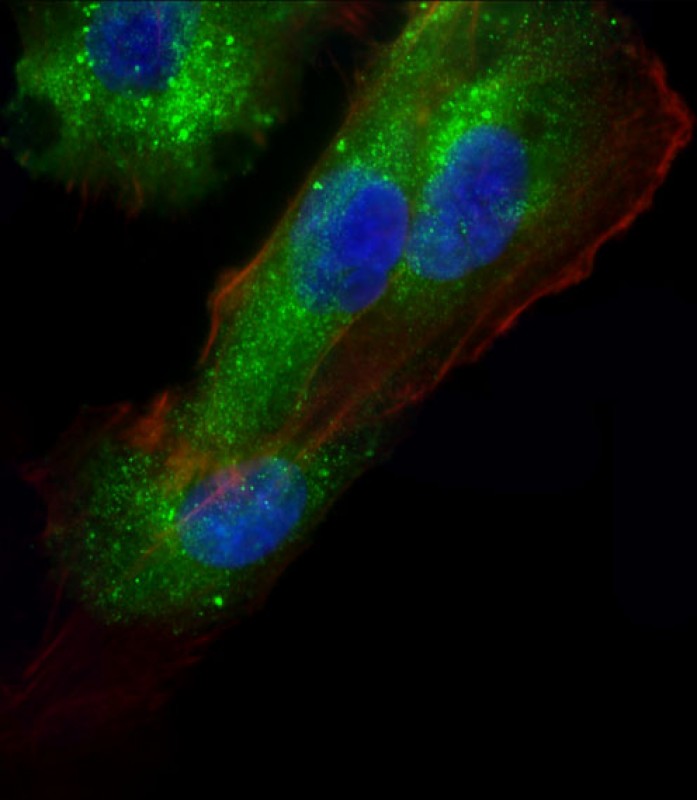 |
Immunofluorescent analysis of 4% paraformaldehyde-fixed, 0. 1% Triton X-100 permeabilized U-251 MG cells labeling AP1M1 with P34586 at 1/25 dilution, followed by Dylight® 488-conjugated goat anti-Rabbit IgG secondary antibody at 1/200 dilution (green). Immunofluorescence image showing Cytoplasm and Weak Nucleus staining on U-251 MG cell line. Cytoplasmic actin is detected with Dylight® 554 Phalloidin(red). The nuclear counter stain is DAPI (blue). |
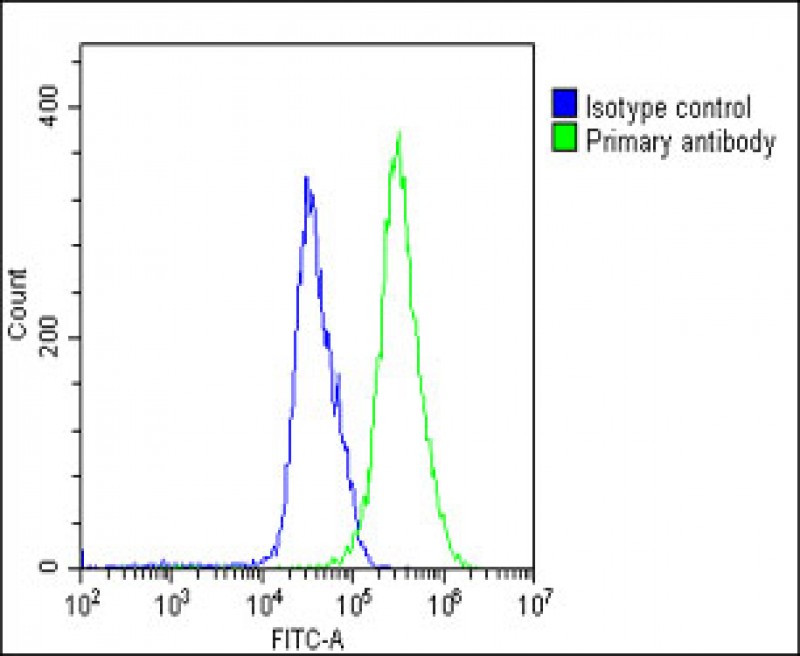 |
Overlay histogram showing U-251 MG cells stained with P34586(green line). The cells were fixed with 2% paraformaldehyde (10 min) and then permeabilized with 90% methanol for 10 min. The cells were then icubated in 2% bovine serum albumin to block non-specific protein-protein interactions followed by the antibody (P34586, 1:25 dilution) for 60 min at 37ºC. The secondary antibody used was Goat-Anti-Rabbit IgG, DyLight® 488 Conjugated Highly Cross-Adsorbed(1583138) at 1/200 dilution for 40 min at 37ºC. Isotype control antibody (blue line) was rabbit IgG1 (1μg/1×10^6 cells) used under the same conditions. Acquisition of >10, 000 events was performed. |
本公司的所有产品仅用于科学研究或者工业应用等非医疗目的,不可用于人类或动物的临床诊断或治疗,非药用,非食用。
暂无评论
本公司的所有产品仅用于科学研究或者工业应用等非医疗目的,不可用于人类或动物的临床诊断或治疗,非药用,非食用。


发表回复