 中文
中文 别名:Actin-related protein 2/3 complex subunit 1A, SOP2-like protein, ARPC1A, SOP2L应用:WB,IHC,ICC,FCM
反应种属:Human
规格:50μl/100μl
| Description |
|---|
| ARPC1A is one of seven subunits of the human Arp2/3 protein complex. This subunit is a member of the SOP2 family of proteins and is most similar to the protein ARPC1B. The similarity between these two proteins suggests that they both may function as p41 subunit of the human Arp2/3 complex that has been implicated in the control of actin polymerization in cells. It is possible that the p41 subunit is involved in assembling and maintaining the structure of the Arp2/3 complex. Multiple versions of the p41 subunit may adapt the functions of the complex to different cell types or developmental stages. |
| Specification | |
|---|---|
| Aliases | Actin-related protein 2/3 complex subunit 1A, SOP2-like protein, ARPC1A, SOP2L |
| Entrez GeneID | 10552 |
| Swissprot | Q92747 |
| WB Predicted band size | 41.6kDa |
| Host/Isotype | Rabbit IgG |
| Storage | Store at 4°C short term. Aliquot and store at -20°C long term. Avoid freeze/thaw cycles. |
| Species Reactivity | Human |
| Immunogen | This ARPC1A antibody is generated from rabbits immunized with a KLH conjugated synthetic peptide between 286-315 amino acids from the C-terminal region of human ARPC1A. |
| Formulation | Purified polyclonal antibody supplied in PBS with 0.05% sodium azide. This antibody is prepared by Saturated Ammonium Sulfate (SAS) precipitation followed by dialysis against PBS. |
| Application | |
|---|---|
| WB | 1/1000 |
| IHC | 1/100-1/500 |
| ICC | 1/10-1/50 |
| FCM | 1/10-1/50 |
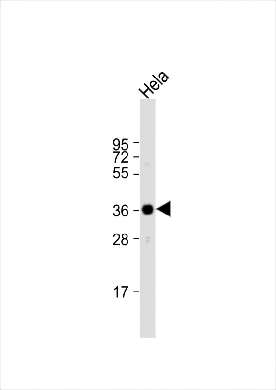 |
Anti-ARPC1A Antibody (C-term) at 1:1000 dilution + Hela whole cell lysate
Lysates/proteins at 20 µg per lane. Secondary Predicted band size : 42 kDa Blocking/Dilution buffer: 5% NFDM/TBST. |
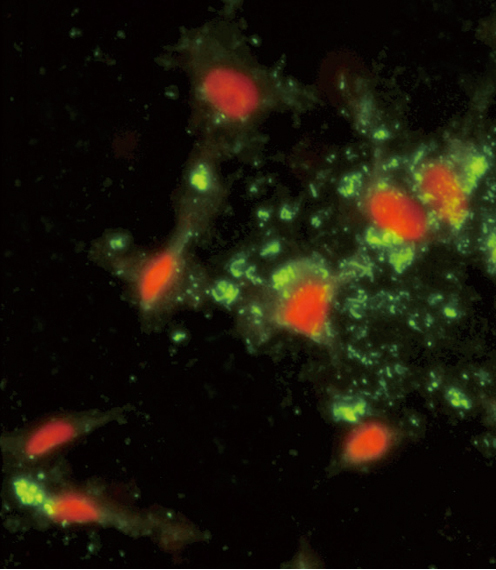 |
Immunofluorescence analysis of ARPC1A Antibody (C-term) with hela cells. 0.025 mg/ml primary antibody was followed by FITC-conjugated goat anti-rabbit lgG (whole molecule). FITC emits green fluorescence.Red counterstaining is PI. |
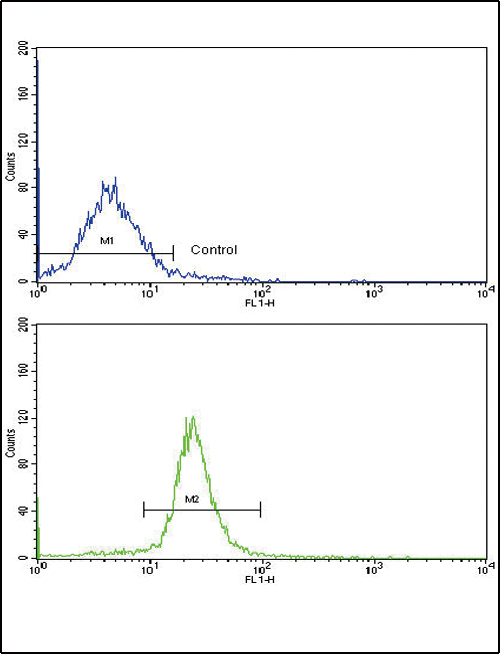 |
Flow cytometric analysis of WiDr cells using ARPC1A Antibody (C-term)(bottom histogram) compared to a negative control cell (top histogram). FITC-conjugated goat-anti-rabbit secondary antibodies were used for the analysis. |
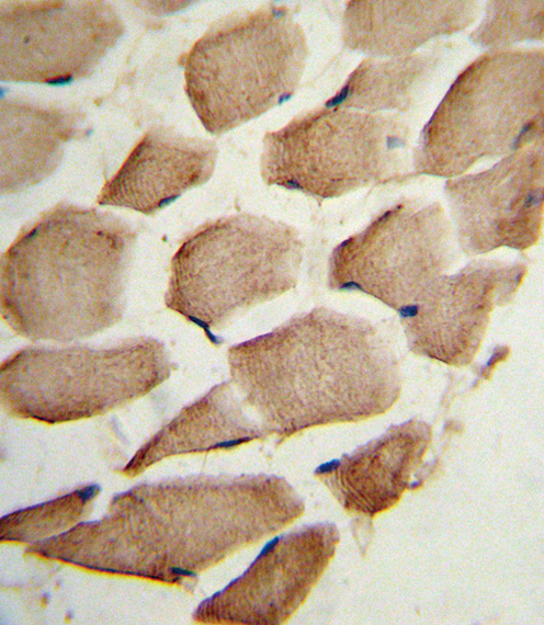 |
Formalin-fixed and paraffin-embedded human skeletal muscle reacted with ARPC1A Antibody (C-term), which was peroxidase-conjugated to the secondary antibody, followed by DAB staining. This data demonstrates the use of this antibody for immunohistochemistry; clinical relevance has not been evaluated. |
本公司的所有产品仅用于科学研究或者工业应用等非医疗目的,不可用于人类或动物的临床诊断或治疗,非药用,非食用。
暂无评论
本公司的所有产品仅用于科学研究或者工业应用等非医疗目的,不可用于人类或动物的临床诊断或治疗,非药用,非食用。


发表回复