 中文
中文 别名:ATP synthase-coupling factor 6, mitochondrial, ATPase subunit F6, ATP5J, ATP5A, ATPM应用:WB,IHC,ICC
反应种属:Human, Mouse, Rat
规格:50μl/100μl
| Description |
|---|
| Mitochondrial ATP synthase catalyzes ATP synthesis, utilizing an electrochemical gradient of protons across the inner membrane during oxidative phosphorylation. It is composed of two linked multi-subunit complexes: the soluble catalytic core, F1, and the membrane-spanning component, Fo, which comprises the proton channel. The F1 complex consists of 5 different subunits (alpha, beta, gamma, delta, and epsilon) assembled in a ratio of 3 alpha, 3 beta, and a single representative of the other 3. The Fo seems to have nine subunits (a, b, c, d, e, f, g, F6 and 8). This gene encodes the F6 subunit of the Fo complex, required for F1 and Fo interactions. Alternatively spliced transcript variants encoding different isoforms have been identified for this gene. A pseudogene exists on chromosome Yp11. |
| Specification | |
|---|---|
| Aliases | ATP synthase-coupling factor 6, mitochondrial, ATPase subunit F6, ATP5J, ATP5A, ATPM |
| Entrez GeneID | 522 |
| Swissprot | P18859 |
| WB Predicted band size | 12.6kDa |
| Host/Isotype | Rabbit IgG |
| Storage | Store at 4°C short term. Aliquot and store at -20°C long term. Avoid freeze/thaw cycles. |
| Species Reactivity | Human, Mouse, Rat |
| Immunogen | This ATP5J antibody is generated from rabbits immunized with a KLH conjugated synthetic peptide between 28-56 amino acids from the Central region of human ATP5J. |
| Formulation | Purified polyclonal antibody supplied in PBS with 0.05% sodium azide. This antibody is purified through a protein A column, followed by peptide affinity purification. |
| Application | |
|---|---|
| WB | 1/1000 |
| IHC | 1/100-1/500 |
| ICC | 1/10-1/50 |
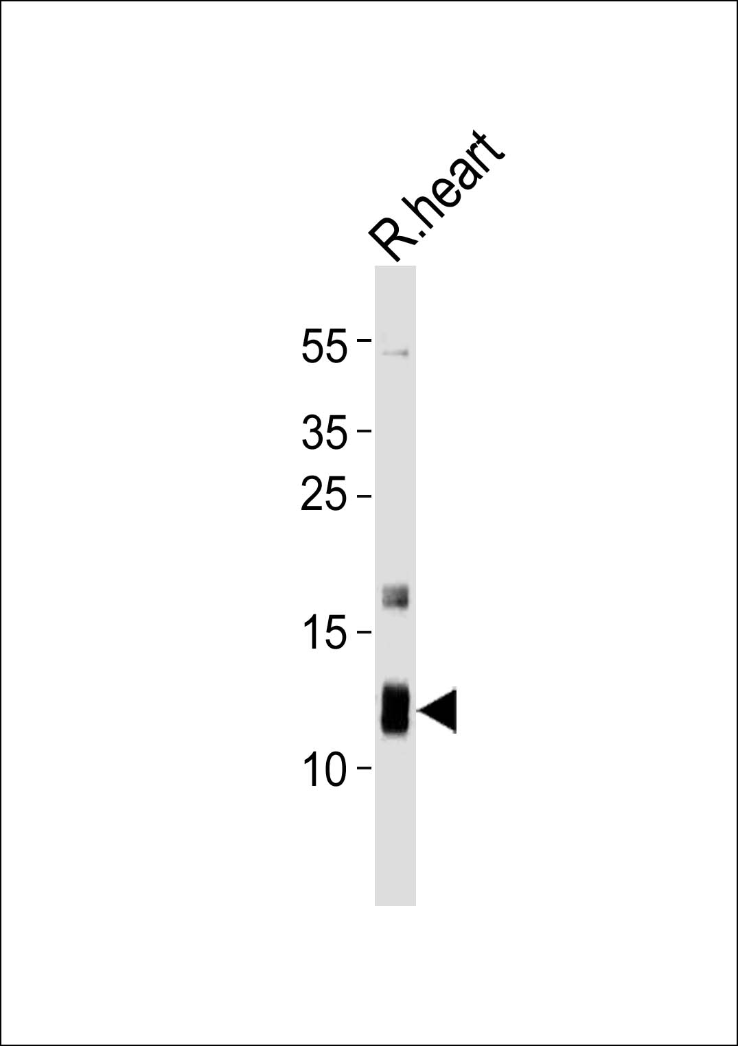 |
Western blot analysis of lysate from rat heart tissue lysate, using ATP5J Antibody (Center)(Cat. #P32785). P32785 was diluted at 1:1000. A goat anti-rabbit IgG H&L(HRP) at 1:10000 dilution was used as the secondary antibody. Lysate at 20ug. |
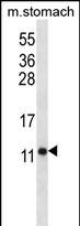 |
ATP5J Antibody (Center) (Cat. #P32785) western blot analysis in mouse stomach tissue lysates (35ug/lane).This demonstrates the ATP5J antibody detected the ATP5J protein (arrow). |
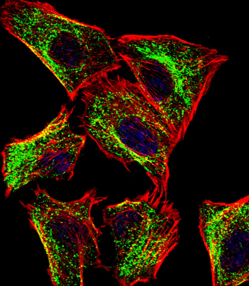 |
Fluorescent confocal image of U251 cell stained with ATP5J Antibody (Center)(Cat#P32785).U251 cells were fixed with 4% PFA (20 min), permeabilized with Triton X-100 (0.1%, 10 min), then incubated with ATP5J primary antibody (1:25, 1 h at 37℃). For secondary antibody, Alexa Fluor® 488 conjugated donkey anti-rabbit antibody (green) was used (1:400, 50 min at 37℃).Cytoplasmic actin was counterstained with Alexa Fluor® 555 (red) conjugated Phalloidin (7units/ml, 1 h at 37℃). Nuclei were counterstained with DAPI (blue) (10 µg/ml, 10 min).ATP5J immunoreactivity is localized to Mitochondria significantly. |
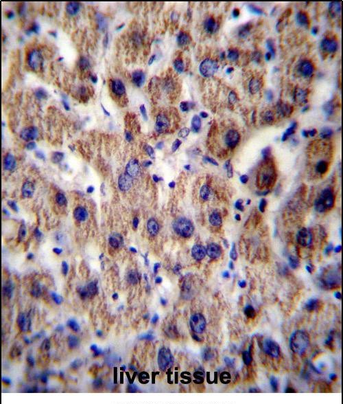 |
ATP5J Antibody (Center) (Cat. #P32785)immunohistochemistry analysis in formalin fixed and paraffin embedded human liver tissue followed by peroxidase conjugation of the secondary antibody and DAB staining.This data demonstrates the use of ATP5J Antibody (Center) for immunohistochemistry. Clinical relevance has not been evaluated. |
本公司的所有产品仅用于科学研究或者工业应用等非医疗目的,不可用于人类或动物的临床诊断或治疗,非药用,非食用。
暂无评论
本公司的所有产品仅用于科学研究或者工业应用等非医疗目的,不可用于人类或动物的临床诊断或治疗,非药用,非食用。


发表回复