 中文
中文 别名:CD59 glycoprotein, 1F5 antigen, 20 kDa homologous restriction factor, HRF-20, HRF20, MAC-inhibitory protein, MAC-IP, MEM43 antigen, Membrane attack complex inhibition factor, MACIF, Membrane inhibitor of reactive lysis, MIRL, Protectin, CD59, CD59, MIC11, MIN1, MIN2, MIN3, MSK21应用:WB,IHC,ICC,FCM
反应种属:Human
规格:50μl/100μl
| Description |
|---|
| Potent inhibitor of the complement membrane attack complex (MAC) action. Acts by binding to the C8 and/or C9 complements of the assembling MAC, thereby preventing incorporation of the multiple copies of C9 required for complete formation of the osmolytic pore. This inhibitor appears to be species-specific. Involved in signal transduction for T-cell activation complexed to a protein tyrosine kinase. |
| Specification | |
|---|---|
| Aliases | CD59 glycoprotein, 1F5 antigen, 20 kDa homologous restriction factor, HRF-20, HRF20, MAC-inhibitory protein, MAC-IP, MEM43 antigen, Membrane attack complex inhibition factor, MACIF, Membrane inhibitor of reactive lysis, MIRL, Protectin, CD59, CD59, MIC11, MIN1, MIN2, MIN3, MSK21 |
| Entrez GeneID | 966 |
| Swissprot | P13987 |
| WB Predicted band size | 14.1kDa |
| Host/Isotype | Rabbit IgG |
| Storage | Store at 4°C short term. Aliquot and store at -20°C long term. Avoid freeze/thaw cycles. |
| Species Reactivity | Human |
| Immunogen | This CD59 antibody is generated from a rabbit immunized with a KLH conjugated synthetic peptide between 74-110 amino acids from the Central region of human CD59. |
| Application | |
|---|---|
| WB | 1/2000 |
| IHC | 1/250-1/1000 |
| ICC | 1/25 |
| FCM | 1/25 |
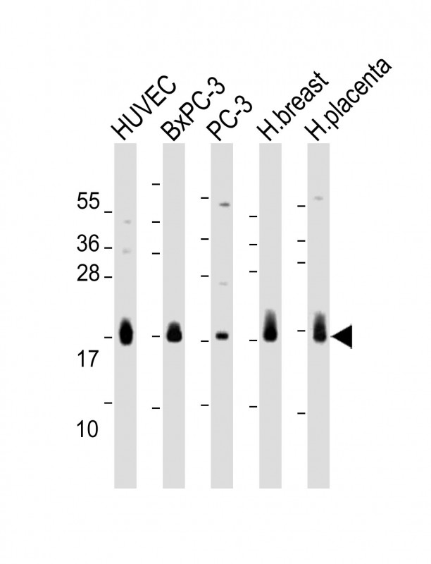 |
All lanes : Anti-CD59 Antibody (Center) at 1:2000 dilution Lane 1: HUVEC whole cell lysate Lane 2: BxPC-3 whole cell lysate Lane 3: PC-3 whole cell lysate Lane 4: Human breast lysate Lane 5: Human placenta lysate Lysates/proteins at 20 µg per lane. Secondary Predicted band size : 14 kDa Blocking/Dilution buffer: 5% NFDM/TBST. |
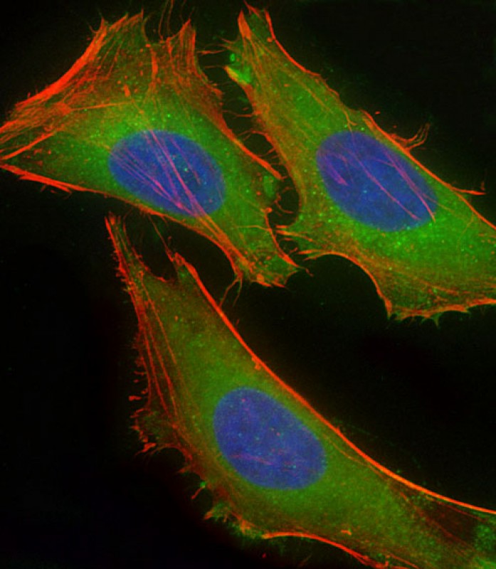 |
Immunofluorescent analysis of 4% paraformaldehyde-fixed, 0.1% Triton X-100 permeabilized HeLa (human cervical epithelial adenocarcinoma cell line) cells labeling CD59 with P34463 at 1/25 dilution, followed by Dylight® 488-conjugated goat anti-rabbit IgG secondary antibody at 1/200 dilution (green). Immunofluorescence image showing cytoplasm staining on HeLa cell line. Cytoplasmic actin is detected with Dylight® 554 Phalloidin at 1/100 dilution (red). The nuclear counter stain is DAPI (blue). |
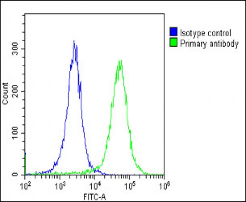 |
Overlay histogram showing HeLa cells stained with P34463(green line). The cells were fixed with 2% paraformaldehyde (10 min) and then permeabilized with 90% methanol for 10 min. The cells were then icubated in 2% bovine serum albumin to block non-specific protein-protein interactions followed by the antibody (P34463, 1:25 dilution) for 60 min at 37ºC. The secondary antibody used was Goat-Anti-Rabbit IgG, DyLight® 488 Conjugated Highly Cross-Adsorbed(OE188374) at 1/200 dilution for 40 min at 37ºC. Isotype control antibody (blue line) was rabbit IgG1 (1μg/1×10^6 cells) used under the same conditions. Acquisition of >10, 000 events was performed. |
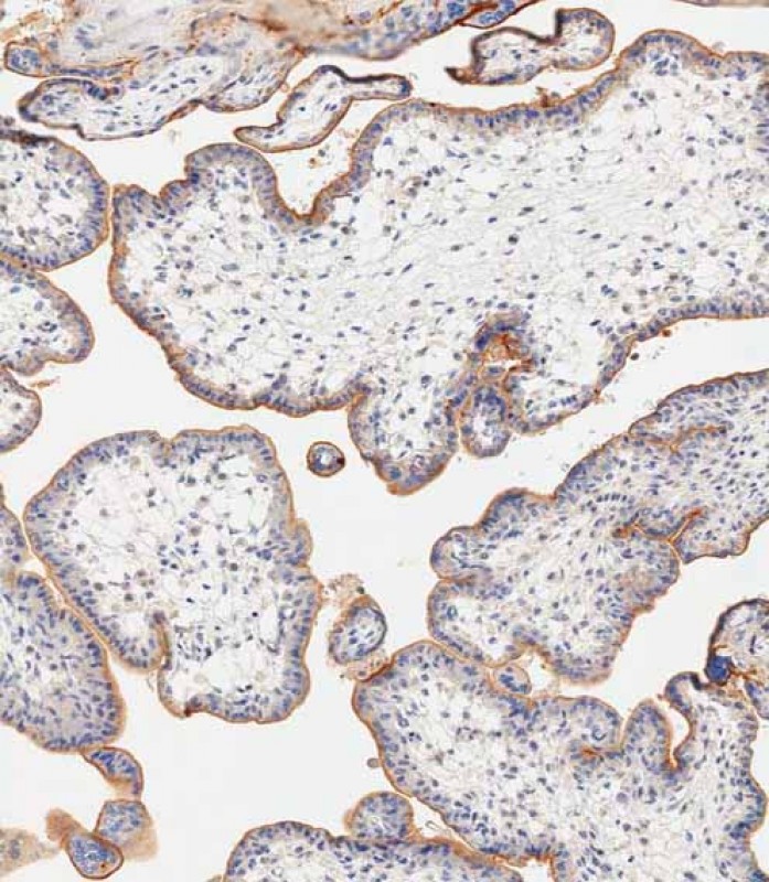 |
Immunohistochemical analysis of paraffin-embedded human placent tissue using P34463 performed on the Leica® BOND RXm. Tissue was fixed with formaldehyde at room temperature; antigen retrieval was by heat mediation with a EDTA buffer (pH9. 0). Samples were incubated with primary antibody(1:1000) for 1 hours at room temperature. A undiluted biotinylated CRF Anti-Polyvalent HRP Polymer antibody was used as the secondary antibody. |
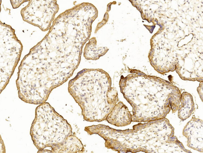 |
Immunohistochemical analysis of paraffin-embedded Human placenta section using Pink1(Cat#P34463). P34463 was diluted at 1:250 dilution. A undiluted biotinylated goat polyvalent antibody was used as the secondary, followed by DAB staining. |
本公司的所有产品仅用于科学研究或者工业应用等非医疗目的,不可用于人类或动物的临床诊断或治疗,非药用,非食用。
暂无评论
本公司的所有产品仅用于科学研究或者工业应用等非医疗目的,不可用于人类或动物的临床诊断或治疗,非药用,非食用。


发表回复