 中文
中文 别名:Cyclin-dependent kinase 4 inhibitor B, Multiple tumor suppressor 2, MTS-2, p14-INK4b, p15-INK4b, p15INK4B, CDKN2B, MTS2应用:WB,IHC,ICC,FCM
反应种属:Human
规格:50μl/100μl
| Description |
|---|
| This gene lies adjacent to the tumor suppressor gene CDKN2A in a region that is frequently mutated and deleted in a wide variety of tumors. This gene encodes a cyclin-dependent kinase inhibitor, which forms a complex with CDK4 or CDK6, and prevents the activation of the CDK kinases, thus the encoded protein functions as a cell growth regulator that controls cell cycle G1 progression. The expression of this gene was found to be dramatically induced by TGF beta, which suggested its role in the TGF beta induced growth inhibition. Two alternatively spliced transcript variants of this gene, which encode distinct proteins, have been reported. |
| Specification | |
|---|---|
| Aliases | Cyclin-dependent kinase 4 inhibitor B, Multiple tumor suppressor 2, MTS-2, p14-INK4b, p15-INK4b, p15INK4B, CDKN2B, MTS2 |
| Entrez GeneID | 1030 |
| Swissprot | P42772 |
| WB Predicted band size | 14.7kDa |
| Host/Isotype | Rabbit IgG |
| Storage | Store at 4°C short term. Aliquot and store at -20°C long term. Avoid freeze/thaw cycles. |
| Species Reactivity | Human |
| Immunogen | This CDKN2B antibody is generated from rabbits immunized with a KLH conjugated synthetic peptide between 103-131 amino acids from the C-terminal region of human CDKN2B. |
| Formulation | Purified polyclonal antibody supplied in PBS with 0.05% sodium azide. This antibody is purified through a protein A column, followed by peptide affinity purification. |
| Application | |
|---|---|
| WB | 1/2000 |
| IHC | 1/100-1/500 |
| ICC | 1/25 |
| FCM | 1/25 |
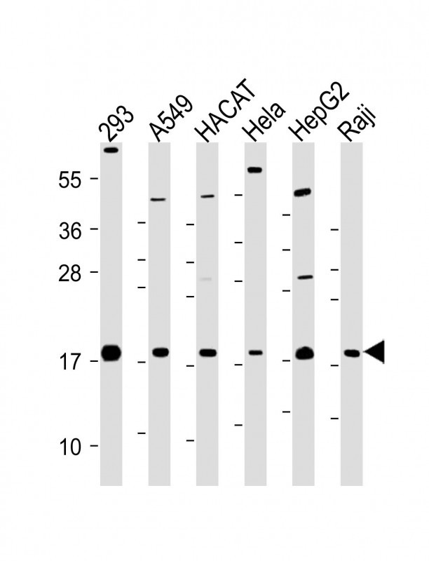 |
All lanes : Anti-CDKN2B Antibody (C-term) at 1:2000 dilution Lane 1: 293 whole cell lysate Lane 2: A549 whole cell lysate Lane 3: HACAT whole cell lysate Lane 4: Hela whole cell lysate Lane 5: HepG2 whole cell lysate Lane 6: Raji whole cell lysate Lysates/proteins at 20 µg per lane. Secondary Predicted band size : 15 kDa Blocking/Dilution buffer: 5% NFDM/TBST. |
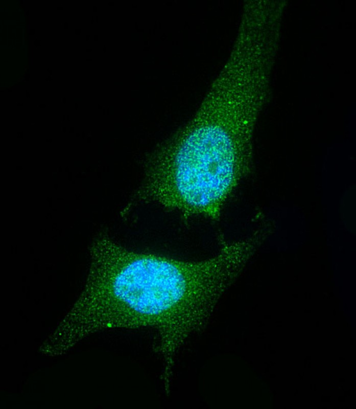 |
Immunofluorescent analysis of 4% paraformaldehyde-fixed, 0. 1% Triton X-100 permeabilized Hela cells labeling CDKN2B with P34459 at 1/25 dilution, followed by Dylight® 488-conjugated goat anti-Rabbit IgG secondary antibody at 1/200 dilution (green). Immunofluorescence image showing Nucleus and Weak Cytoplasm staining on Hela cell line. The nuclear counter stain is DAPI (blue). |
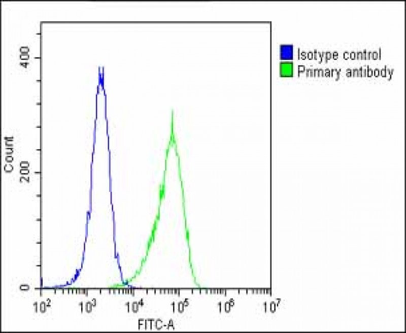 |
Overlay histogram showing Hela cells stained with P34459(green line). The cells were fixed with 2% paraformaldehyde (10 min) and then permeabilized with 90% methanol for 10 min. The cells were then icubated in 2% bovine serum albumin to block non-specific protein-protein interactions followed by the antibody (P34459, 1:25 dilution) for 60 min at 37ºC. The secondary antibody used was Goat-Anti-Rabbit IgG, DyLight® 488 Conjugated Highly Cross-Adsorbed(1583138) at 1/200 dilution for 40 min at 37ºC. Isotype control antibody (blue line) was rabbit IgG1 (1μg/1×10^6 cells) used under the same conditions. Acquisition of >10, 000 events was performed. |
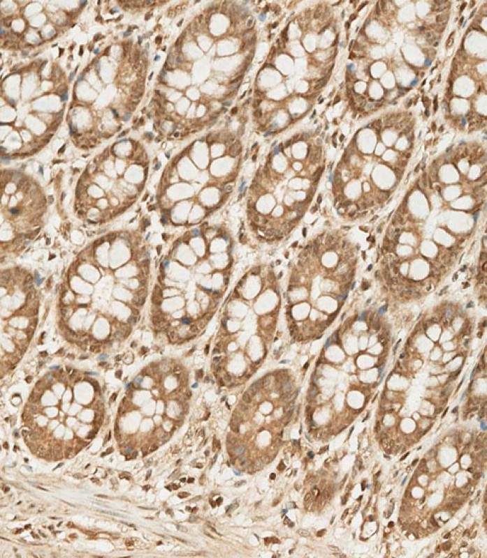 |
P34459 staining CDKN2B in human colon tissue sections by Immunohistochemistry (IHC-P – paraformaldehyde-fixed, paraffin-embedded sections). Samples were incubated with primary antibody (1/250) for 1 hours at room temperature. A undiluted biotinylated goat polyvalent antibody was used as the secondary antibody. |
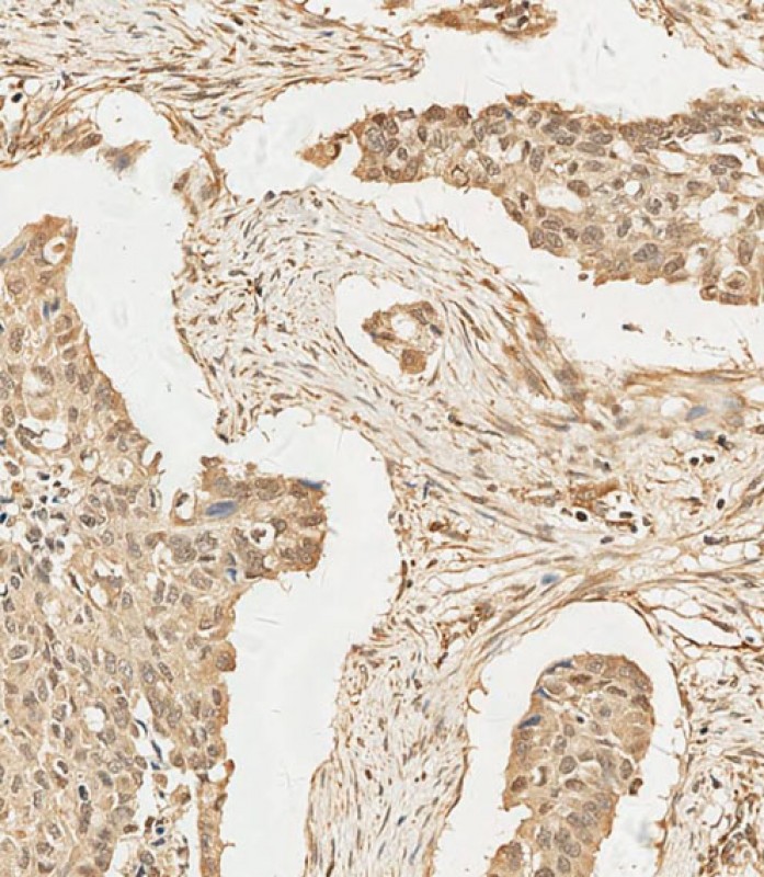 |
P34459 staining CDKN2B in human lung adenocarcinoma tissue sections by Immunohistochemistry (IHC-P – paraformaldehyde-fixed, paraffin-embedded sections). Samples were incubated with primary antibody (1/100) for 1 hours at room temperature. A undiluted biotinylated goat polyvalent antibody was used as the secondary antibody. |
本公司的所有产品仅用于科学研究或者工业应用等非医疗目的,不可用于人类或动物的临床诊断或治疗,非药用,非食用。
暂无评论
本公司的所有产品仅用于科学研究或者工业应用等非医疗目的,不可用于人类或动物的临床诊断或治疗,非药用,非食用。


发表回复