 中文
中文 别名:Collagen alpha-1(XIV) chain, Undulin, COL14A1 (HGNC:2191), UND应用:WB,IHC,FCM
反应种属:Human, Mouse
规格:50μl/100μl
| Description |
|---|
| Plays an adhesive role by integrating collagen bundles. It is probably associated with the surface of interstitial collagen fibrils via COL1. The COL2 domain may then serve as a rigid arm which sticks out from the fibril and protrudes the large N-terminal globular domain into the extracellular space, where it might interact with other matrix molecules or cell surface receptors (By similarity). |
| Specification | |
|---|---|
| Aliases | Collagen alpha-1(XIV) chain, Undulin, COL14A1 (HGNC:2191), UND |
| Entrez GeneID | 7373 |
| Swissprot | Q05707 |
| WB Predicted band size | 193.5kDa |
| Host/Isotype | Rabbit IgG |
| Storage | Store at 4°C short term. Aliquot and store at -20°C long term. Avoid freeze/thaw cycles. |
| Species Reactivity | Human, Mouse |
| Immunogen | This COL14A1 antibody is generated from a rabbit immunized with a KLH conjugated synthetic peptide between 643-677 amino acids from the Central region of human COL14A1. |
| Application | |
|---|---|
| WB | 1/2000 |
| IHC | 1/100-1/500 |
| FCM | 1/25 |
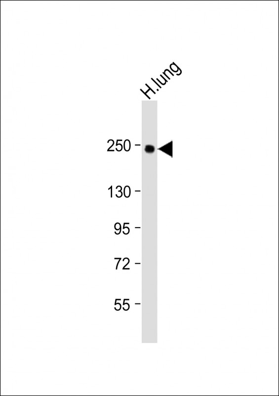 |
Anti-COL14A1 Antibody (Center) at 1:2000 dilution + Human lung lysate
Lysates/proteins at 20 µg per lane. Secondary Predicted band size : 194 kDa Blocking/Dilution buffer: 5% NFDM/TBST. |
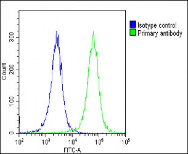 |
Overlay histogram showing Hela cells stained with P34514(green line). The cells were fixed with 2% paraformaldehyde (10 min) and then permeabilized with 90% methanol for 10 min. The cells were then icubated in 2% bovine serum albumin to block non-specific protein-protein interactions followed by the antibody (P34514, 1:25 dilution) for 60 min at 37ºC. The secondary antibody used was Goat-Anti-Rabbit IgG, DyLight® 488 Conjugated Highly Cross-Adsorbed(OE188374) at 1/200 dilution for 40 min at 37ºC. Isotype control antibody (blue line) was rabbit IgG1 (1μg/1×10^6 cells) used under the same conditions. Acquisition of >10, 000 events was performed. |
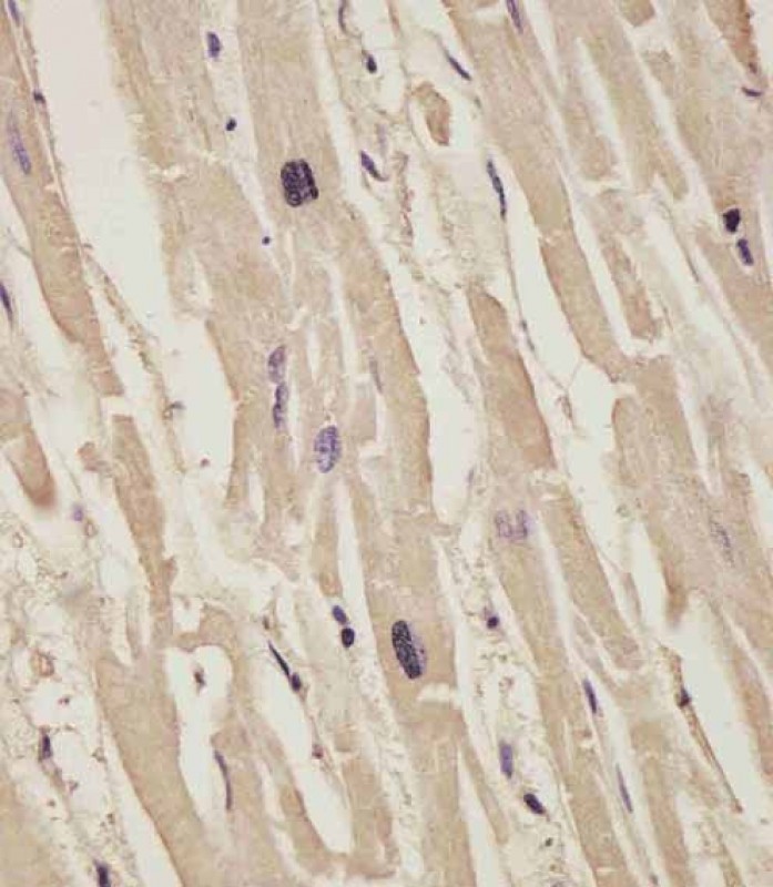 |
P34514 staining COL14A1 in human heart tissue sections by Immunohistochemistry (IHC-P – paraformaldehyde-fixed, paraffin-embedded sections). Tissue was fixed with formaldehyde and blocked with 3% BSA for 0. 5 hour at room temperature; antigen retrieval was by heat mediation with a citrate buffer (pH6). Samples were incubated with primary antibody (1/25) for 1 hours at 37°C. A undiluted biotinylated goat polyvalent antibody was used as the secondary antibody. |
本公司的所有产品仅用于科学研究或者工业应用等非医疗目的,不可用于人类或动物的临床诊断或治疗,非药用,非食用。
暂无评论
本公司的所有产品仅用于科学研究或者工业应用等非医疗目的,不可用于人类或动物的临床诊断或治疗,非药用,非食用。


发表回复