 中文
中文 别名:COP9 signalosome complex subunit 7b, SGN7b, Signalosome subunit 7b, JAB1-containing signalosome subunit 7b, COPS7B, CSN7B应用:WB,IHC,FCM
反应种属:Human, Mouse
规格:50μl/100μl
| Description |
|---|
| Component of the COP9 signalosome complex (CSN), a complex involved in various cellular and developmental processes. The CSN complex is an essential regulator of the ubiquitin (Ubl) conjugation pathway by mediating the deneddylation of the cullin subunits of SCF-type E3 ligase complexes, leading to decrease the Ubl ligase activity of SCF-type complexes such as SCF, CSA or DDB2. The complex is also involved in phosphorylation of p53/TP53, JUN, I-kappa-B-alpha/NFKBIA, ITPK1 and IRF8/ICSBP, possibly via its association with CK2 and PKD kinases. CSN-dependent phosphorylation of TP53 and JUN promotes and protects degradation by the Ubl system, respectively. |
| Specification | |
|---|---|
| Aliases | COP9 signalosome complex subunit 7b, SGN7b, Signalosome subunit 7b, JAB1-containing signalosome subunit 7b, COPS7B, CSN7B |
| Entrez GeneID | 64708 |
| Swissprot | Q9H9Q2 |
| WB Predicted band size | 29.6kDa |
| Host/Isotype | Rabbit IgG |
| Storage | Store at 4°C short term. Aliquot and store at -20°C long term. Avoid freeze/thaw cycles. |
| Species Reactivity | Human, Mouse |
| Immunogen | This COPS7B antibody is generated from a rabbit immunized with a KLH conjugated synthetic peptide between 61-95 amino acids from human COPS7B. |
| Application | |
|---|---|
| WB | 1/2000 |
| IHC | 1/100-1/500 |
| FCM | 1/25 |
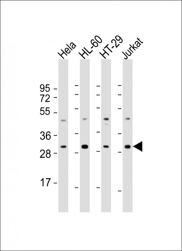 |
All lanes : Anti-COPS7B Antibody (N-Term) at 1:2000 dilution Lane 1: Hela whole cell lysate Lane 2: HL-60 whole cell lysate Lane 3: HT-29 whole cell lysate Lane 4: Jurkat whole cell lysate Lysates/proteins at 20 µg per lane. Secondary Predicted band size : 30 kDa Blocking/Dilution buffer: 5% NFDM/TBST. |
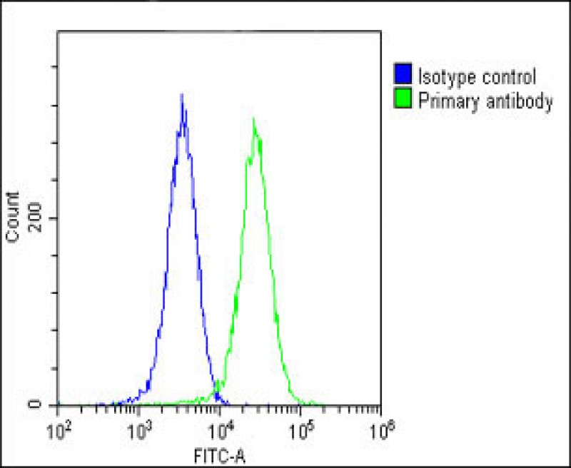 |
Overlay histogram showing Hela cells stained with P34289(green line). The cells were fixed with 2% paraformaldehyde (10 min) and then permeabilized with 90% methanol for 10 min. The cells were then icubated in 2% bovine serum albumin to block non-specific protein-protein interactions followed by the antibody (P34289, 1:25 dilution) for 60 min at 37ºC. The secondary antibody used was Goat-Anti-Rabbit IgG, DyLight® 488 Conjugated Highly Cross-Adsorbed(OH191631) at 1/200 dilution for 40 min at 37ºC. Isotype control antibody (blue line) was rabbit IgG1 (1μg/1×10^6 cells) used under the same conditions. Acquisition of >10, 000 events was performed. |
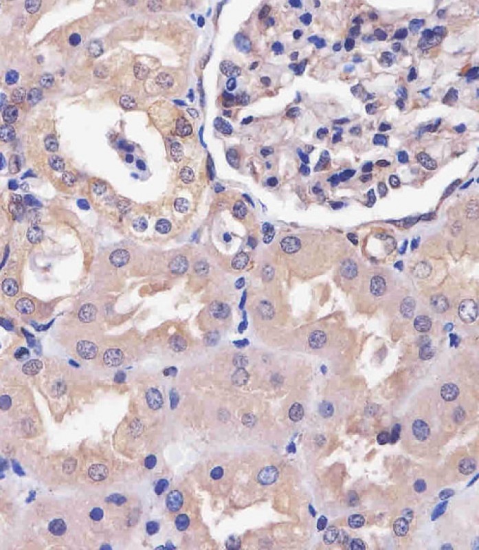 |
P34289 staining COPS7B in human kidney tissue sections by Immunohistochemistry (IHC-P – paraformaldehyde-fixed, paraffin-embedded sections). Tissue was fixed with formaldehyde and blocked with 3% BSA for 0. 5 hour at room temperature; antigen retrieval was by heat mediation with a citrate buffer (pH6). Samples were incubated with primary antibody (1/25) for 1 hours at 37°C. A undiluted biotinylated goat polyvalent antibody was used as the secondary antibody. |
本公司的所有产品仅用于科学研究或者工业应用等非医疗目的,不可用于人类或动物的临床诊断或治疗,非药用,非食用。
暂无评论
本公司的所有产品仅用于科学研究或者工业应用等非医疗目的,不可用于人类或动物的临床诊断或治疗,非药用,非食用。


发表回复