 中文
中文 别名:Cyclic AMP-responsive element-binding protein 1, CREB-1, cAMP-responsive element-binding protein 1, CREB1应用:WB,IHC,ICC
反应种属:Human
规格:50μl/100μl
| Description |
|---|
| Phosphorylation-dependent transcription factor that stimulates transcription upon binding to the DNA cAMP response element (CRE), a sequence present in many viral and cellular promoters. Transcription activation is enhanced by the TORC coactivators which act independently of Ser-133 phosphorylation. Involved in different cellular processes including the synchronization of circadian rhythmicity and the differentiation of adipose cells. |
| Specification | |
|---|---|
| Aliases | Cyclic AMP-responsive element-binding protein 1, CREB-1, cAMP-responsive element-binding protein 1, CREB1 |
| Entrez GeneID | 1385 |
| Swissprot | P16220 |
| WB Predicted band size | 35.1kDa |
| Host/Isotype | Rabbit IgG |
| Storage | Store at 4°C short term. Aliquot and store at -20°C long term. Avoid freeze/thaw cycles. |
| Species Reactivity | Human |
| Immunogen | This CREB(S133) antibody is generated from a rabbit immunized with a KLH conjugated synthetic peptide between 110-140 from the human region of human CREB(S133). |
| Application | |
|---|---|
| WB | 1/2000 |
| IHC | 1/500 |
| ICC | 1/25 |
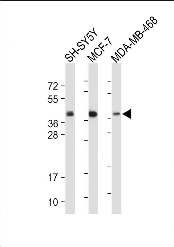 |
All lanes : Anti-CREB(S133) Antibody at 1:2000 dilution Lane 1: SH-SY5Y whole cell lysate Lane 2: MCF-7 whole cell lysate Lane 3: MDA-MB-468 whole cell lysate Lysates/proteins at 20 µg per lane. Secondary Predicted band size : 37 kDa Blocking/Dilution buffer: 5% NFDM/TBST. |
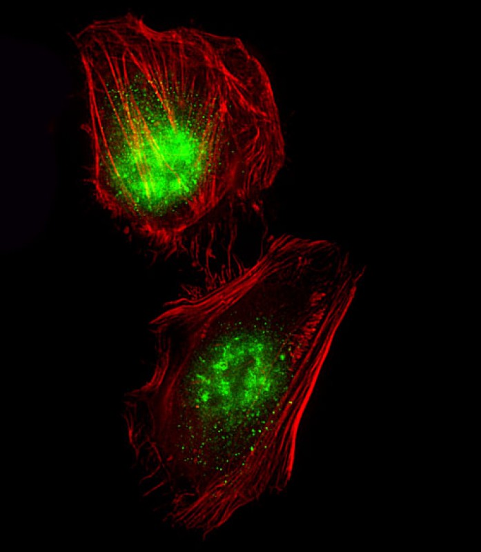 |
Immunofluorescent analysis of 4% paraformaldehyde-fixed, 0. 1% Triton X-100 permeabilized Hela cells labeling CREB1 with P34708 at 1/25 dilution, followed by Dylight® 488-conjugated goat anti-Rabbit IgG secondary antibody at 1/200 dilution (green). Immunofluorescence image showing Nucleus and Weak Cytoplasm staining on Hela cell line. Cytoplasmic actin is detected with Dylight® 554 Phalloidin(red). The nuclear counter stain is DAPI (blue). |
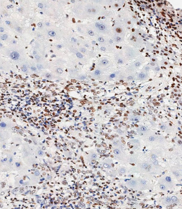 |
Immunohistochemical analysis of paraffin-embedded Human hepatocarcinoma tissue using P34708 performed on the Leica® BOND RXm. Tissue was fixed with formaldehyde at room temperature, antigen retrieval was by heat mediation with a EDTA buffer (pH9. 0). Samples were incubated with primary antibody(1:500) for 1 hours at room temperature. A undiluted biotinylated CRF Anti-Polyvalent HRP Polymer antibody was used as the secondary antibody. |
本公司的所有产品仅用于科学研究或者工业应用等非医疗目的,不可用于人类或动物的临床诊断或治疗,非药用,非食用。
暂无评论
本公司的所有产品仅用于科学研究或者工业应用等非医疗目的,不可用于人类或动物的临床诊断或治疗,非药用,非食用。


发表回复