 中文
中文 别名:Transcription factor CP2-like protein 1, CP2-related transcriptional repressor 1, CRTR-1, Transcription factor LBP-9, TFCP2L1, CRTR1, LBP9应用:WB,IHC,ICC,FCM
反应种属:Human, Mouse, Rat
规格:50μl/100μl
| Description |
|---|
| Transcriptional suppressor. CRTRT1 may suppress UBP1-mediated transcriptional activation. Modulates the placental expression of CYP11A1. |
| Specification | |
|---|---|
| Aliases | Transcription factor CP2-like protein 1, CP2-related transcriptional repressor 1, CRTR-1, Transcription factor LBP-9, TFCP2L1, CRTR1, LBP9 |
| Entrez GeneID | 29842 |
| Swissprot | Q9NZI6 |
| WB Predicted band size | 54.6kDa |
| Host/Isotype | Rabbit IgG |
| Storage | Store at 4°C short term. Aliquot and store at -20°C long term. Avoid freeze/thaw cycles. |
| Species Reactivity | Human, Mouse, Rat |
| Immunogen | This CRTR1 antibody is generated from rabbits immunized with a KLH conjugated synthetic peptide between 14-44 amino acids from the N-terminal region of human CRTR1. |
| Formulation | Purified polyclonal antibody supplied in PBS with 0.05% sodium azide. This antibody is purified through a protein A column, followed by peptide affinity purification. |
| Application | |
|---|---|
| WB | 1/2000 |
| IHC | 1/500 |
| ICC | 1/25 |
| FCM | 1/25 |
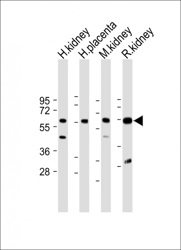 |
All lanes : Anti-CRTRT1 Antibody (N-term) at 1:2000 dilution Lane 1: Human kidney lysate Lane 2: Human placenta lysate Lane 3: Mouse kidney lysate Lane 4: Rat kidney lysate Lysates/proteins at 20 µg per lane. Secondary Predicted band size : 55 kDa Blocking/Dilution buffer: 5% NFDM/TBST. |
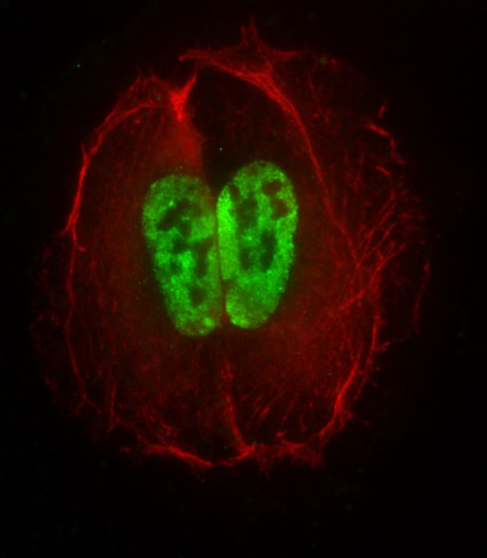 |
Immunofluorescent analysis of 4% paraformaldehyde-fixed, 0. 1% Triton X-100 permeabilized U-2OS cells labeling TFCP2L1 with P34531 at 1/25 dilution, followed by Dylight® 488-conjugated goat anti-Rabbit IgG secondary antibody at 1/200 dilution (green). Immunofluorescence image showing nucleus staining on U-2OS cell line. Cytoplasmic actin is detected with Dylight® 554 Phalloidin at 1/500 dilution (red). The nuclear counter stain is DAPI (blue). |
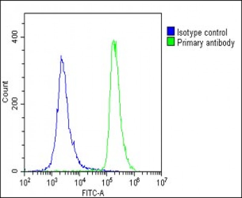 |
Overlay histogram showing U-2 OS cells stained with P34531(green line). The cells were fixed with 2% paraformaldehyde (10 min) and then permeabilized with 90% methanol for 10 min. The cells were then icubated in 2% bovine serum albumin to block non-specific protein-protein interactions followed by the antibody (P34531, 1:25 dilution) for 60 min at 37ºC. The secondary antibody used was Goat-Anti-Rabbit IgG, DyLight® 488 Conjugated Highly Cross-Adsorbed(1583138) at 1/200 dilution for 40 min at 37ºC. Isotype control antibody (blue line) was rabbit IgG1 (1μg/1×10^6 cells) used under the same conditions. Acquisition of >10, 000 events was performed. |
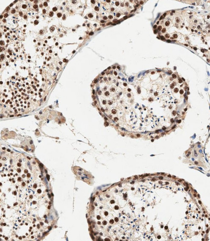 |
Immunohistochemical analysis of paraffin-embedded human testis tissue using P34531 performed on the Leica® BOND RXm. Samples were incubated with primary antibody(1/500) for 1 hours at room temperature. A undiluted biotinylated CRF Anti-Polyvalent HRP Polymer antibody was used as the secondary antibody. |
本公司的所有产品仅用于科学研究或者工业应用等非医疗目的,不可用于人类或动物的临床诊断或治疗,非药用,非食用。
暂无评论
本公司的所有产品仅用于科学研究或者工业应用等非医疗目的,不可用于人类或动物的临床诊断或治疗,非药用,非食用。


发表回复