 中文
中文 别名:Dopamine beta-hydroxylase, Dopamine beta-monooxygenase, Soluble dopamine beta-hydroxylase, DBH应用:WB,ICC
反应种属:Human, Mouse, Rat
规格:50μl/100μl
| Description |
|---|
| The protein encoded by this gene is an oxidoreductase belonging to the copper type II, ascorbate-dependent monooxygenase family. It is present in the synaptic vesicles of postganglionic sympathetic neurons and converts dopamine to norepinephrine. It exists in both soluble and membrane-bound forms, depending on the absence or presence, respectively, of a signal peptide. [provided by RefSeq]. |
| Specification | |
|---|---|
| Aliases | Dopamine beta-hydroxylase, Dopamine beta-monooxygenase, Soluble dopamine beta-hydroxylase, DBH |
| Entrez GeneID | 1621 |
| Swissprot | P09172 |
| WB Predicted band size | 69.1kDa |
| Host/Isotype | Rabbit IgG |
| Storage | Store at 4°C short term. Aliquot and store at -20°C long term. Avoid freeze/thaw cycles. |
| Species Reactivity | Human, Mouse, Rat |
| Immunogen | This DBH antibody is generated from rabbits immunized with a KLH conjugated synthetic peptide between 27-56 amino acids from the N-terminal region of human DBH. |
| Formulation | Purified polyclonal antibody supplied in PBS with 0.05% sodium azide. This antibody is prepared by Saturated Ammonium Sulfate (SAS) precipitation followed by dialysis against PBS. |
| Application | |
|---|---|
| WB | 1/2000 |
| ICC | 1/25 |
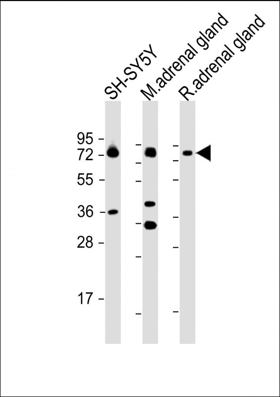 |
All lanes : Anti-DBH Antibody (N-term P42) at 1:2000 dilution Lane 1: SH-SY5Y whole cell lysate Lane 2: mouse adrenal gland lysate Lane 3: rat adrenal gland lysate Lysates/proteins at 20 µg per lane. Secondary Predicted band size : 69 kDa Blocking/Dilution buffer: 5% NFDM/TBST. |
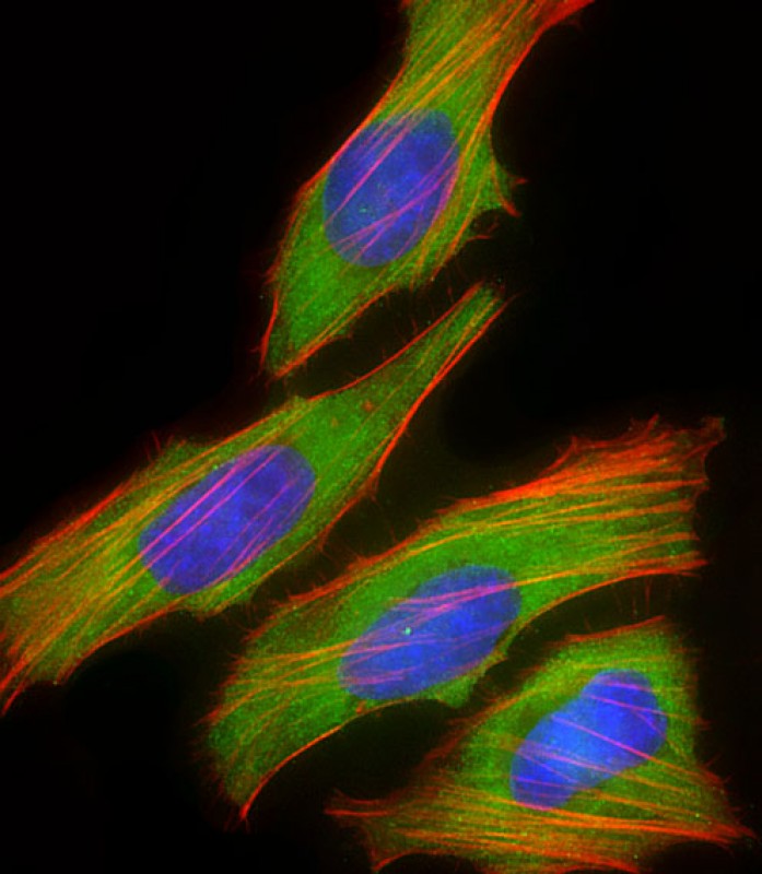 |
Immunofluorescent analysis of 4% paraformaldehyde-fixed, 0.1% Triton X-100 permeabilized HeLa (human cervical epithelial adenocarcinoma cell line) cells labeling DBH with P34386 at 1/25 dilution, followed by Dylight® 488-conjugated goat anti-rabbit IgG secondary antibody at 1/200 dilution (green). Immunofluorescence image showing cytoplasm and weak nucleus staining on HeLa cell line. Cytoplasmic actin is detected with Dylight® 554 Phalloidin at 1/100 dilution (red).The nuclear counter stain is DAPI (blue). |
本公司的所有产品仅用于科学研究或者工业应用等非医疗目的,不可用于人类或动物的临床诊断或治疗,非药用,非食用。
暂无评论
本公司的所有产品仅用于科学研究或者工业应用等非医疗目的,不可用于人类或动物的临床诊断或治疗,非药用,非食用。


发表回复