 中文
中文 别名:Putative elongation factor 1-alpha-like 3, EF-1-alpha-like 3, Eukaryotic elongation factor 1 A-like 3, eEF1A-like 3, Eukaryotic translation elongation factor 1 alpha-1 pseudogene 5, EEF1A1P5, EEF1AL3应用:WB,ICC,FCM
反应种属:Human
规格:50μl/100μl
| Description |
|---|
| This protein promotes the GTP-dependent binding of aminoacyl-tRNA to the A-site of ribosomes during protein biosynthesis. |
| Specification | |
|---|---|
| Aliases | Putative elongation factor 1-alpha-like 3, EF-1-alpha-like 3, Eukaryotic elongation factor 1 A-like 3, eEF1A-like 3, Eukaryotic translation elongation factor 1 alpha-1 pseudogene 5, EEF1A1P5, EEF1AL3 |
| Swissprot | Q5VTE0 |
| WB Predicted band size | 50.2kDa |
| Host/Isotype | Rabbit IgG |
| Storage | Store at 4°C short term. Aliquot and store at -20°C long term. Avoid freeze/thaw cycles. |
| Species Reactivity | Human |
| Immunogen | This EEF1A1P5 antibody is generated from a rabbit immunized with a KLH conjugated synthetic peptide between 430-462 amino acids from human EEF1A1P5. |
| Application | |
|---|---|
| WB | 1/1000-1/2000 |
| ICC | 1/25 |
| FCM | 1/25 |
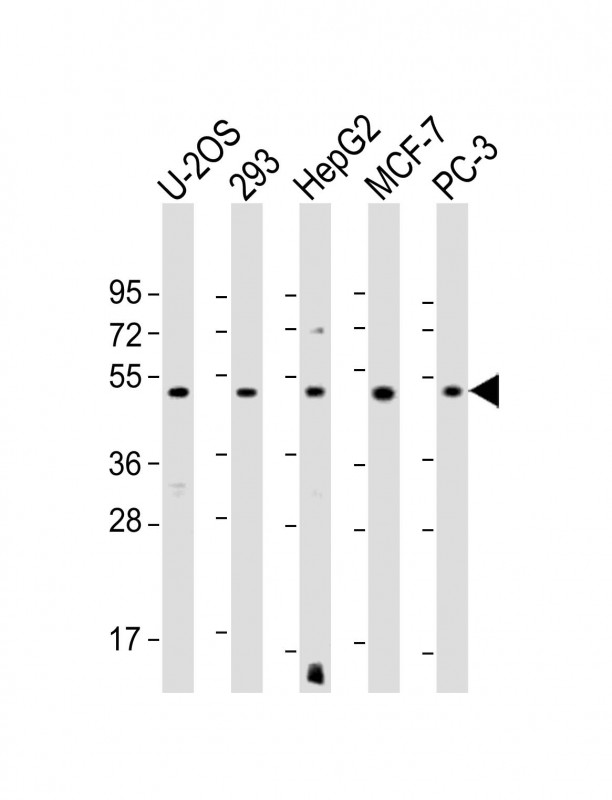 |
All lanes : Anti-EEF1A1P5 Antibody (C-Term) at 1:1000-1:2000 dilution Lane 1: U-2OS whole cell lysate Lane 2: 293 whole cell lysate Lane 3: HepG2 whole cell lysate Lane 4: MCF-7 whole cell lysate Lane 5: PC-3 whole cell lysate Lysates/proteins at 20 µg per lane. Secondary Predicted band size : 50 kDa Blocking/Dilution buffer: 5% NFDM/TBST. |
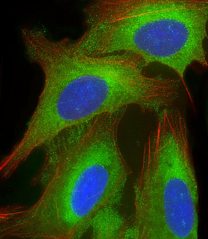 |
Immunofluorescent analysis of 4% paraformaldehyde-fixed, 0.1% Triton X-100 permeabilized U-2 OS (human osteosarcoma cell line) cells labeling EEF1A1P5 with P34361 at 1/25 dilution, followed by Dylight® 488-conjugated goat anti-rabbit IgG secondary antibody at 1/200 dilution (green). Immunofluorescence image showing cytoplasm staining on U-2 OS cell line. Cytoplasmic actin is detected with Dylight® 554 Phalloidin at 1/100 dilution (red).The nuclear counter stain is DAPI (blue). |
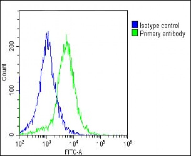 |
Overlay histogram showing HepG2 cells stained with P34361(green line). The cells were fixed with 2% paraformaldehyde (10 min) and then permeabilized with 90% methanol for 10 min. The cells were then icubated in 2% bovine serum albumin to block non-specific protein-protein interactions followed by the antibody (P34361, 1:25 dilution) for 60 min at 37ºC. The secondary antibody used was Goat-Anti-Rabbit IgG, DyLight® 488 Conjugated Highly Cross-Adsorbed(OE188374) at 1/200 dilution for 40 min at 37ºC. Isotype control antibody (blue line) was rabbit IgG1 (1μg/1×10^6 cells) used under the same conditions. Acquisition of >10, 000 events was performed. . |
本公司的所有产品仅用于科学研究或者工业应用等非医疗目的,不可用于人类或动物的临床诊断或治疗,非药用,非食用。
暂无评论
本公司的所有产品仅用于科学研究或者工业应用等非医疗目的,不可用于人类或动物的临床诊断或治疗,非药用,非食用。


发表回复