 中文
中文 别名:Equilibrative nucleoside transporter 1, Equilibrative nitrobenzylmercaptopurine riboside-sensitive nucleoside transporter, Equilibrative NBMPR-sensitive nucleoside transporter, Nucleoside transporter, es-type, Solute carrier family 29 member 1, SLC29A1, ENT1应用:WB,IHC,FCM
反应种属:Human, Mouse, Rat
规格:50μl/100μl
| Description |
|---|
| ENT1 is a member of the equilibrative nucleoside transporter family. It is a transmembrane glycoprotein that localizes to the plasma and mitochondrial membranes and mediates the cellular uptake of nucleosides from the surrounding medium. The protein is categorized as an equilibrative (as opposed to concentrative) transporter that is sensitive to inhibition by nitrobenzylthioinosine (NBMPR). Nucleoside transporters are required for nucleotide synthesis in cells that lack de novo nucleoside synthesis pathways, and are also necessary for the uptake of cytotoxic nucleosides used for cancer and viral chemotherapies. |
| Specification | |
|---|---|
| Aliases | Equilibrative nucleoside transporter 1, Equilibrative nitrobenzylmercaptopurine riboside-sensitive nucleoside transporter, Equilibrative NBMPR-sensitive nucleoside transporter, Nucleoside transporter, es-type, Solute carrier family 29 member 1, SLC29A1, ENT1 |
| Entrez GeneID | 2030 |
| Swissprot | Q99808 |
| WB Predicted band size | 50.2kDa |
| Host/Isotype | Rabbit IgG |
| Storage | Store at 4°C short term. Aliquot and store at -20°C long term. Avoid freeze/thaw cycles. |
| Species Reactivity | Human, Mouse, Rat |
| Immunogen | This ENT1 antibody is generated from rabbits immunized with a KLH conjugated synthetic peptide between 402-431 amino acids from the C-terminal region of human ENT1. |
| Formulation | Purified polyclonal antibody supplied in PBS with 0.05% sodium azide. This antibody is purified through a protein A column, followed by peptide affinity purification. |
| Application | |
|---|---|
| WB | 1/1000 |
| IHC | 1/500 |
| FCM | 1/25 |
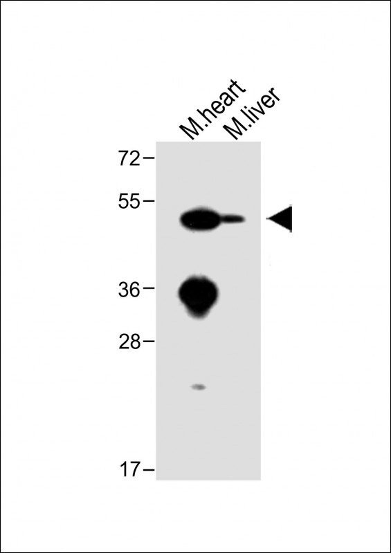 |
All lanes : Anti-ENT1 Antibody (C-term) at 1:1000 dilution Lane 1: Mouse heart lysate Lane 2: Mouse liver lysate Lysates/proteins at 20 µg per lane. Secondary Predicted band size : 50 kDa Blocking/Dilution buffer: 5% NFDM/TBST. |
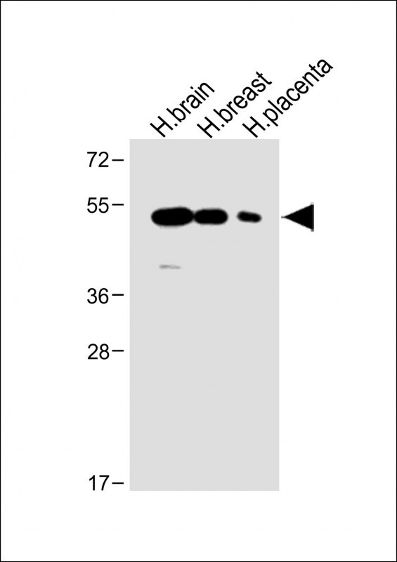 |
All lanes : Anti-ENT1 Antibody (C-term) at 1:1000 dilution Lane 1: Human brain lysate Lane 2: Human breast lysate Lane 3: Human placenta lysate Lysates/proteins at 20 µg per lane. Secondary Predicted band size : 50 kDa Blocking/Dilution buffer: 5% NFDM/TBST. |
 |
Western blot analysis of lysate from human heart tissue lysate, using ENT1(Slc29a1) Antibody (C-term)(Cat. #P34585). P34585 was diluted at 1:1000. A goat anti-rabbit IgG H&L(HRP) at 1:5000 dilution was used as the secondary antibody. Lysate at 35ug. |
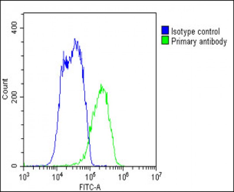 |
Overlay histogram showing HepG2 cells stained with P34585(green line). The cells were fixed with 2% paraformaldehyde 10 min. The cells were then icubated in 2% bovine serum albumin to block non-specific protein-protein interactions followed by the antibody (P34585, 1:25 dilution) for 60 min at 37ºC. The secondary antibody used was Goat-Anti-Rabbit IgG, DyLight® 488 Conjugated Highly Cross-Adsorbed(1583138) at 1/200 dilution for 40 min at 37ºC. Isotype control antibody (blue line) was rabbit IgG1 (1μg/1×10^6 cells) used under the same conditions. Acquisition of >10, 000 events was performed. |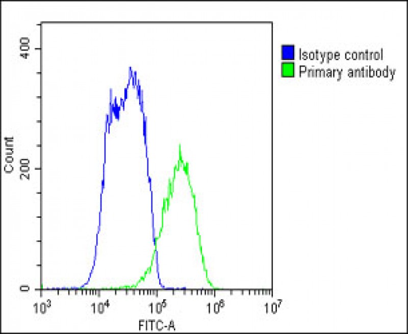
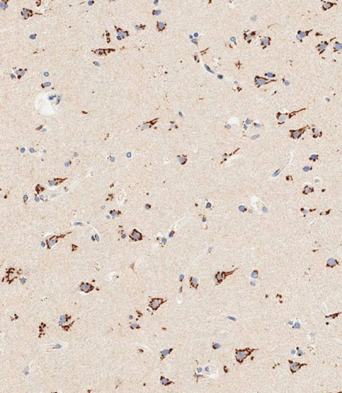 |
Immunohistochemical analysis of paraffin-embedded human brain tissue using P34585 performed on the Leica® BOND RXm. Samples were incubated with primary antibody(1/500) for 1 hours at room temperature. A undiluted biotinylated CRF Anti-Polyvalent HRP Polymer antibody was used as the secondary antibody. |
本公司的所有产品仅用于科学研究或者工业应用等非医疗目的,不可用于人类或动物的临床诊断或治疗,非药用,非食用。
暂无评论
本公司的所有产品仅用于科学研究或者工业应用等非医疗目的,不可用于人类或动物的临床诊断或治疗,非药用,非食用。


发表回复