 中文
中文 别名:Ephrin type-A receptor 2, Epithelial cell kinase, Tyrosine-protein kinase receptor ECK, EPHA2, ECK应用:WB,IHC,FCM
反应种属:Human, Mouse
规格:50μl/100μl
| Description |
|---|
| Protein kinases are enzymes that transfer a phosphate group from a phosphate donor, generally the g phosphate of ATP, onto an acceptor amino acid in a substrate protein. By this basic mechanism, protein kinases mediate most of the signal transduction in eukaryotic cells, regulating cellular metabolism, transcription, cell cycle progression, cytoskeletal rearrangement and cell movement, apoptosis, and differentiation. With more than 500 gene products, the protein kinase family is one of the largest families of proteins in eukaryotes. The family has been classified in 8 major groups based on sequence comparison of their tyrosine (PTK) or serine/threonine (STK) kinase catalytic domains. The tyrosine kinase (TK) group is mainly involved in the regulation of cell-cell interactions such as differentiation, adhesion, motility and death. There are currently about 90 TK genes sequenced, 58 are of receptor protein TK (e.g. EGFR, EPH, FGFR, PDGFR, TRK, and VEGFR families), and 32 of cytosolic TK (e.g. ABL, FAK, JAK, and SRC families). |
| Specification | |
|---|---|
| Aliases | Ephrin type-A receptor 2, Epithelial cell kinase, Tyrosine-protein kinase receptor ECK, EPHA2, ECK |
| Entrez GeneID | 1969 |
| Swissprot | P29317 |
| WB Predicted band size | 108.3kDa |
| Host/Isotype | Rabbit IgG |
| Storage | Store at 4°C short term. Aliquot and store at -20°C long term. Avoid freeze/thaw cycles. |
| Species Reactivity | Human, Mouse |
| Immunogen | This EphA2 antibody is generated from rabbits immunized with a KLH conjugated synthetic peptide between 30-60 amino acids from the N-terminal region of human EphA2. |
| Formulation | Purified polyclonal antibody supplied in PBS with 0.05% sodium azide. This antibody is purified through a protein G column, eluted with high and low pH buffers and neutralized immediately, followed by dialysis against PBS. |
| Application | |
|---|---|
| WB | 1/1000 |
| IHC | 1/100-1/500 |
| FCM | 1/10-1/50 |
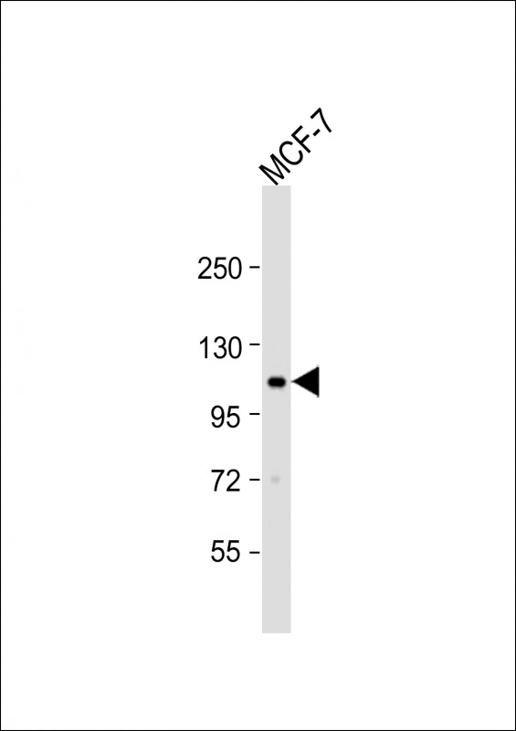 |
Anti-EPHA2 Antibody (T45) at 1:1000 dilution + MCF-7 whole cell lysate
Lysates/proteins at 20 µg per lane. Secondary Predicted band size : 108 kDa Blocking/Dilution buffer: 5% NFDM/TBST. |
 |
Western blot analysis of hEPHA2-T45 (Cat. #P33578) in MCF-7 cell line lysates (35ug/lane). EPHA2 (arrow) was detected using the purified Pab |
 |
Western blot analysis of hEPHA2-T45 (Cat. #P33578) in mouse NIH-3T3 cell line lysates (35ug/lane). EPHA2 (arrow) was detected using the purified Pab. |
 |
Flow cytometric analysis of NCI-H292 cells using EphA2 Antibody (N-term)(bottom histogram) compared to a negative control cell (top histogram). FITC-conjugated goat-anti-rabbit secondary antibodies were used for the analysis. |
 |
Formalin-fixed and paraffin-embedded human cancer tissue reacted with the primary antibody, which was peroxidase-conjugated to the secondary antibody, followed by AEC staining. This data demonstrates the use of this antibody for immunohistochemistry; clinical relevance has not been evaluated. BC = breast carcinoma; HC = hepatocarcinoma. |
本公司的所有产品仅用于科学研究或者工业应用等非医疗目的,不可用于人类或动物的临床诊断或治疗,非药用,非食用。
暂无评论
本公司的所有产品仅用于科学研究或者工业应用等非医疗目的,不可用于人类或动物的临床诊断或治疗,非药用,非食用。


发表回复