 中文
中文 别名:Growth factor receptor-bound protein 14, GRB14 adapter protein, GRB14应用:WB,ICC,FCM
反应种属:Human, Mouse, Rat
规格:50μl/100μl
| Description |
|---|
| Adapter protein which modulates coupling of cell surface receptor kinases with specific signaling pathways. Binds to, and suppresses signals from, the activated insulin receptor (INSR). Potent inhibitor of insulin-stimulated MAPK3 phosphorylation. Plays a critical role regulating PDPK1 membrane translocation in response to insulin stimulation and serves as an adapter protein to recruit PDPK1 to activated insulin receptor, thus promoting PKB/AKT1 phosphorylation and transduction of the insulin signal. |
| Specification | |
|---|---|
| Aliases | Growth factor receptor-bound protein 14, GRB14 adapter protein, GRB14 |
| Entrez GeneID | 2888 |
| Swissprot | Q14449 |
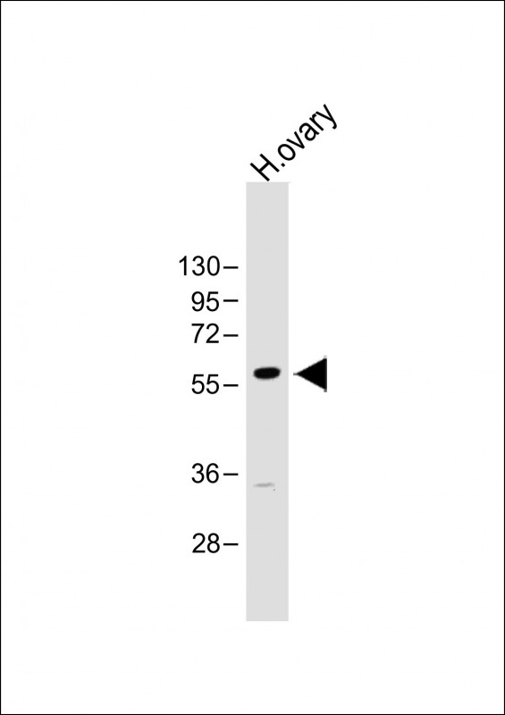
| WB Predicted band size | 61.0kDa |
| Host/Isotype | Rabbit IgG |
| Storage | Store at 4°C short term. Aliquot and store at -20°C long term. Avoid freeze/thaw cycles. |
| Species Reactivity | Human, Mouse, Rat |
| Immunogen | This GRB14 antibody is generated from a rabbit immunized with a KLH conjugated synthetic peptide between 14-48 amino acids from the human region of human GRB14. |
| Formulation | Purified polyclonal antibody supplied in PBS with 0.05% sodium azide. This antibody is purified through a protein A column, followed by peptide affinity purification. |
| Application | |
|---|---|
| WB | 1/1000 |
| ICC | 1/25 |
| FCM | 1/25 |
 |
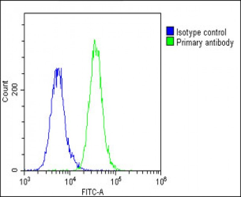
Overlay histogram showing A549 cells stained with P34644(green line). The cells were fixed with 2% paraformaldehyde (10 min) and then permeabilized with 90% methanol for 10 min. The cells were then icubated in 2% bovine serum albumin to block non-specific protein-protein interactions followed by the antibody (P34644, 1:25 dilution) for 60 min at 37ºC.
The secondary antibody used was Goat-Anti-Rabbit IgG,DyLight® 488 Conjugated Highly Cross-Adsorbed(1583138) at 1/200 dilution for 40 min at 37ºC. Isotype control antibody (blue line) was rabbit IgG1 (1μg/1×10^6 cells) used under the same conditions. Acquisition of >10,000 events was performed. |
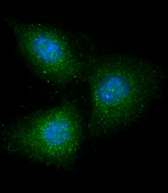 |
Immunofluorescent analysis of 4% paraformaldehyde-fixed, 0. 1% Triton X-100 permeabilized A549 cells labeling GRB14 with P34644 at 1/25 dilution, followed by Dylight® 488-conjugated goat anti-Rabbit IgG secondary antibody at 1/200 dilution (green). Immunofluorescence image showing cytoplasm staining on A549 cell line. Cytoplasmic actin is detected with Dylight® 554 Phalloidin at 1/500 dilution (red). The nuclear counter stain is DAPI (blue). |
 |
Overlay histogram showing A549 cells stained with P34644(green line). The cells were fixed with 2% paraformaldehyde (10 min) and then permeabilized with 90% methanol for 10 min. The cells were then icubated in 2% bovine serum albumin to block non-specific protein-protein interactions followed by the antibody (P34644, 1:25 dilution) for 60 min at 37ºC. The secondary antibody used was Goat-Anti-Rabbit IgG, DyLight® 488 Conjugated Highly Cross-Adsorbed(1583138) at 1/200 dilution for 40 min at 37ºC. Isotype control antibody (blue line) was rabbit IgG1 (1μg/1×10^6 cells) used under the same conditions. Acquisition of >10, 000 events was performed. |
本公司的所有产品仅用于科学研究或者工业应用等非医疗目的,不可用于人类或动物的临床诊断或治疗,非药用,非食用。
暂无评论
本公司的所有产品仅用于科学研究或者工业应用等非医疗目的,不可用于人类或动物的临床诊断或治疗,非药用,非食用。


发表回复