 中文
中文 别名:Histone H2B type 1-J, Histone H2B1, Histone H2Br, H2B/r, HIST1H2BJ, H2BFR应用:WB,IHC,FCM
反应种属:Human
规格:50μl/100μl
| Description |
|---|
| Histones are basic nuclear proteins that are responsible for the nucleosome structure of the chromosomal fiber in eukaryotes. Two molecules of each of the four core histones (H2A, H2B, H3, and H4) form an octamer, around which approximately 146 bp of DNA is wrapped in repeating units, called nucleosomes. The linker histone, H1, interacts with linker DNA between nucleosomes and functions in the compaction of chromatin into higher order structures. This gene is intronless and encodes a member of the histone H2B family. Transcripts from this gene lack polyA tails but instead contain a palindromic termination element. This gene is found in the histone microcluster on chromosome 6p21.33. [provided by RefSeq]. |
| Specification | |
|---|---|
| Aliases | Histone H2B type 1-J, Histone H2B1, Histone H2Br, H2B/r, HIST1H2BJ, H2BFR |
| Entrez GeneID | 8970 |
| Swissprot | P06899 |
| WB Predicted band size | 13.9kDa |
| Host/Isotype | Rabbit IgG |
| Storage | Store at 4°C short term. Aliquot and store at -20°C long term. Avoid freeze/thaw cycles. |
| Species Reactivity | Human |
| Immunogen | This HIST1H2B antibody is generated from rabbits immunized with a KLH conjugated synthetic peptide between 57-86 amino acids from the Central region of human HIST1H2BJ. |
| Formulation | Purified polyclonal antibody supplied in PBS with 0.05% sodium azide. This antibody is purified through a protein A column, followed by peptide affinity purification. |
| Application | |
|---|---|
| WB | 1/1000 |
| IHC | 1/100-1/500 |
| FCM | 1/10-1/50 |
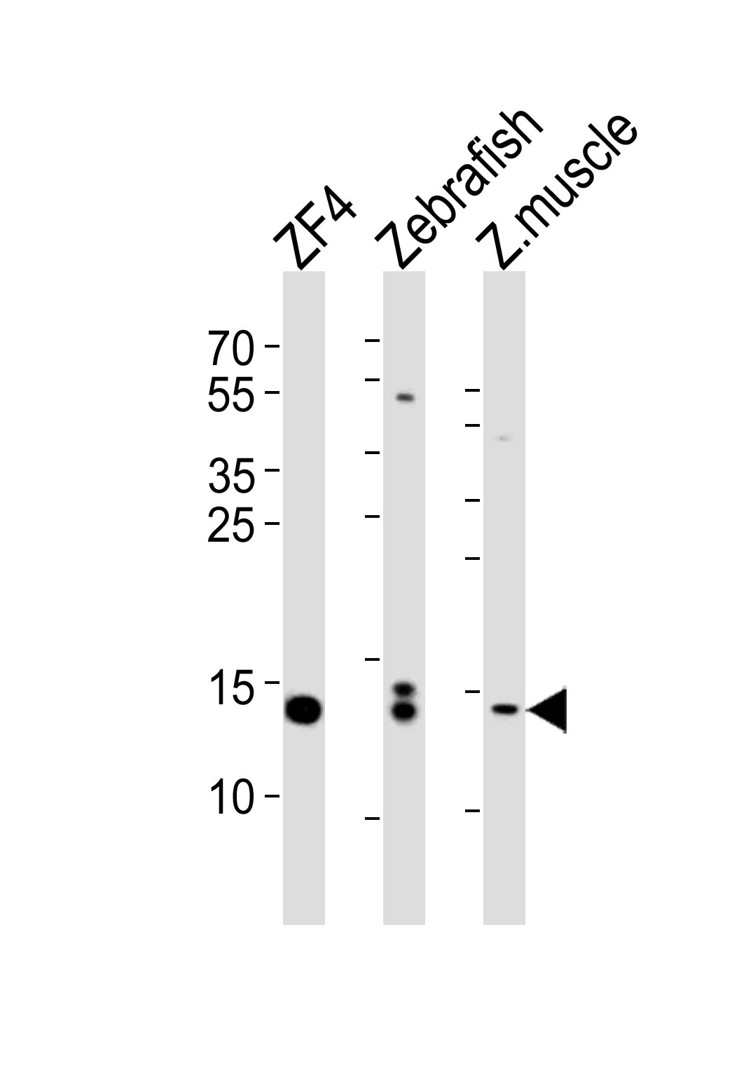 |
Western blot analysis of lysates from ZF4 cell line, Zebrafish, zebra fish muscle tissue lysate(from left to right), using HIST1H2BJ Antibody (Center)(Cat. #P32705). P32705 was diluted at 1:1000 at each lane. A goat anti-rabbit IgG H&L(HRP) at 1:5000 dilution was used as the secondary antibody. Lysates at 35ug per lane. |
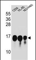 |
HIST1H2BJ Antibody (Center) (Cat. #P32705) western blot analysis in CEM,HL-60,NCI-H460 cell line lysates (35ug/lane).This demonstrates the HIST1H2BJ antibody detected the HIST1H2BJ protein (arrow). |
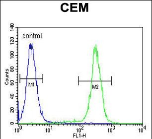 |
HIST1H2BJ Antibody (Center) (Cat. #P32705) flow cytometric analysis of CEM cells (right histogram) compared to a negative control cell (left histogram).FITC-conjugated goat-anti-rabbit secondary antibodies were used for the analysis. |
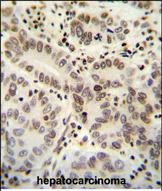 |
HIST1H2BJ antibody (Center) (Cat. #P32705) immunohistochemistry analysis in formalin fixed and paraffin embedded human hepatocarcinoma followed by peroxidase conjugation of the secondary antibody and DAB staining. This data demonstrates the use of the HIST1H2BJ antibody (Center) for immunohistochemistry. Clinical relevance has not been evaluated. |
本公司的所有产品仅用于科学研究或者工业应用等非医疗目的,不可用于人类或动物的临床诊断或治疗,非药用,非食用。
暂无评论
本公司的所有产品仅用于科学研究或者工业应用等非医疗目的,不可用于人类或动物的临床诊断或治疗,非药用,非食用。


发表回复