 中文
中文 别名:HLA class II histocompatibility antigen, DP beta 1 chain, HLA class II histocompatibility antigen, DP(W4) beta chain, MHC class II antigen DPB1, HLA-DPB1, HLA-DP1B应用:WB,IHC,FCM
反应种属:Human
规格:50μl/100μl
| Description |
|---|
| HLA-DPB belongs to the HLA class II beta chain paralogues. This class II molecule is a heterodimer consisting of an alpha (DPA) and a beta chain (DPB), both anchored in the membrane. It plays a central role in the immune system by presenting peptides derived from extracellular proteins. Class II molecules are expressed in antigen presenting cells (APC: B lymphocytes, dendritic cells, macrophages). The beta chain is approximately 26-28 kDa and its gene contains 6 exons. Exon one encodes the leader peptide, exons 2 and 3 encode the two extracellular domains, exon 4 encodes the transmembrane domain and exon 5 encodes the cytoplasmic tail. Within the DP molecule both the alpha chain and the beta chain contain the polymorphisms specifying the peptide binding specificities, resulting in up to 4 different molecules. |
| Specification | |
|---|---|
| Aliases | HLA class II histocompatibility antigen, DP beta 1 chain, HLA class II histocompatibility antigen, DP(W4) beta chain, MHC class II antigen DPB1, HLA-DPB1, HLA-DP1B |
| Entrez GeneID | 3115 |
| Swissprot | P04440 |
| WB Predicted band size | 29.2kDa |
| Host/Isotype | Rabbit IgG |
| Storage | Store at 4°C short term. Aliquot and store at -20°C long term. Avoid freeze/thaw cycles. |
| Species Reactivity | Human |
| Immunogen | This HLA-DPB1 antibody is generated from rabbits immunized with a KLH conjugated synthetic peptide between 77-105 amino acids from the Central region of human HLA-DPB1. |
| Formulation | Purified polyclonal antibody supplied in PBS with 0.05% sodium azide. This antibody is purified through a protein A column, followed by peptide affinity purification. |
| Application | |
|---|---|
| WB | 1/1000 |
| IHC | 1/500 |
| FCM | 1/25 |
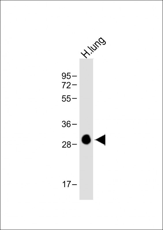 |
Anti-HLA-DPB1 Antibody (Center) at 1:1000 dilution + Human lung lysate
Lysates/proteins at 20 µg per lane. Secondary Predicted band size : 30 kDa Blocking/Dilution buffer: 5% NFDM/TBST. |
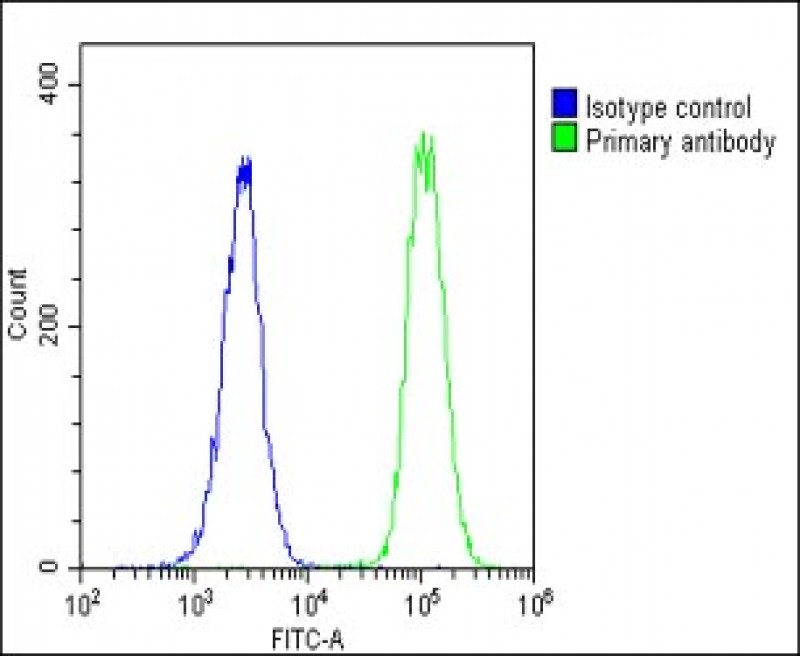 |
Overlay histogram showing U-2 OS cells stained with P34577(green line). The cells were fixed with 2% paraformaldehyde (10 min) and then permeabilized with 90% methanol for 10 min. The cells were then icubated in 2% bovine serum albumin to block non-specific protein-protein interactions followed by the antibody (P34577, 1:25 dilution) for 60 min at 37ºC. The secondary antibody used was Goat-Anti-Rabbit IgG, DyLight® 488 Conjugated Highly Cross-Adsorbed(1583138) at 1/200 dilution for 40 min at 37ºC. Isotype control antibody (blue line) was rabbit IgG1 (1μg/1×10^6 cells) used under the same conditions. Acquisition of >10, 000 events was performed. |
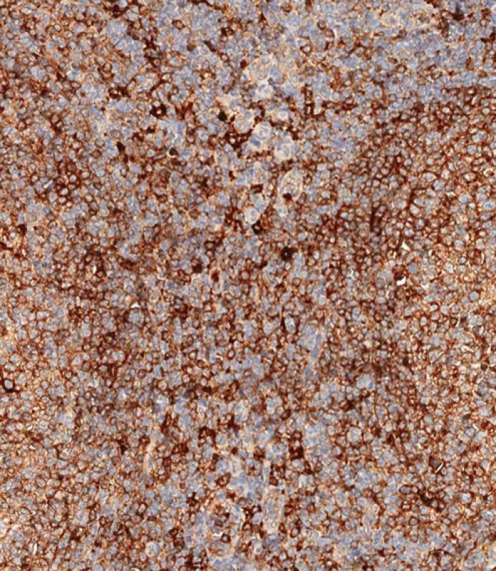 |
Immunohistochemical analysis of paraffin-embedded human tonsil tissue using P34577 performed on the Leica® BOND RXm. Samples were incubated with primary antibody(1/500) for 1 hours at room temperature. A undiluted biotinylated CRF Anti-Polyvalent HRP Polymer antibody was used as the secondary antibody. |
本公司的所有产品仅用于科学研究或者工业应用等非医疗目的,不可用于人类或动物的临床诊断或治疗,非药用,非食用。
暂无评论
本公司的所有产品仅用于科学研究或者工业应用等非医疗目的,不可用于人类或动物的临床诊断或治疗,非药用,非食用。


发表回复