 中文
中文 别名:3-hydroxyacyl-CoA dehydrogenase type-2, 1.1.1.35, 17-beta-hydroxysteroid dehydrogenase 10, 17-beta-HSD 10, 1.1.1.51, 3-hydroxy-2-methylbutyryl-CoA dehydrogenase, 1.1.1.178, 3-hydroxyacyl-CoA dehydrogenase type II, Endoplasmic reticulum-associated amyloid beta-peptide-binding protein, Mitochondrial ribonuclease P protein 2, Mitochondrial RNase P protein 2, Short-chain type dehydrogenase/reductase XH98G2, Type II HADH, HSD17B10, ERAB, HADH2, MRPP2, SCHAD, XH98G2应用:WB,IHC,ICC,FCM
反应种属:Human, Mouse, Rat
规格:50μl/100μl
| Description |
|---|
| Functions in mitochondrial tRNA maturation. Part of mitochondrial ribonuclease P, an enzyme composed of MRPP1/TRMT10C, MRPP2/HSD17B10 and MRPP3/KIAA0391, which cleaves tRNA molecules in their 5′-ends. Catalyzes the beta-oxidation at position 17 of androgens and estrogens and has 3-alpha-hydroxysteroid dehydrogenase activity with androsterone. Catalyzes the third step in the beta-oxidation of fatty acids. Carries out oxidative conversions of 7-alpha-OH and 7-beta-OH bile acids. Also exhibits 20-beta-OH and 21-OH dehydrogenase activities with C21 steroids. By interacting with intracellular amyloid-beta, it may contribute to the neuronal dysfunction associated with Alzheimer disease (AD). |
| Specification | |
|---|---|
| Aliases | 3-hydroxyacyl-CoA dehydrogenase type-2, 1.1.1.35, 17-beta-hydroxysteroid dehydrogenase 10, 17-beta-HSD 10, 1.1.1.51, 3-hydroxy-2-methylbutyryl-CoA dehydrogenase, 1.1.1.178, 3-hydroxyacyl-CoA dehydrogenase type II, Endoplasmic reticulum-associated amyloid beta-peptide-binding protein, Mitochondrial ribonuclease P protein 2, Mitochondrial RNase P protein 2, Short-chain type dehydrogenase/reductase XH98G2, Type II HADH, HSD17B10, ERAB, HADH2, MRPP2, SCHAD, XH98G2 |
| Entrez GeneID | 3028 |
| Swissprot | Q99714 |
| WB Predicted band size | 26.9kDa |
| Host/Isotype | Rabbit IgG |
| Storage | Store at 4°C short term. Aliquot and store at -20°C long term. Avoid freeze/thaw cycles. |
| Species Reactivity | Human, Mouse, Rat |
| Immunogen | This HSD17B10 antibody is generated from a rabbit immunized with a KLH conjugated synthetic peptide between 140-172 amino acids from the Central region of human HSD17B10. |
| Application | |
|---|---|
| WB | 1/2000 |
| IHC | 1/100-1/500 |
| ICC | 1/25 |
| FCM | 1/25 |
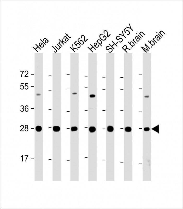 |
All lanes : Anti-HSD17B10 Antibody (Center) at 1:2000 dilution Lane 1: Hela whole cell lysate Lane 2: Jurkat whole cell lysate Lane 3: K562 whole cell lysate Lane 4: HepG2 whole cell lysate Lane 5: SH-SY5Y whole cell lysate Lane 6: rat brain lysate Lane 7: mouse brain lysate Lysates/proteins at 20 µg per lane. Secondary Predicted band size : 27 kDa Blocking/Dilution buffer: 5% NFDM/TBST. |
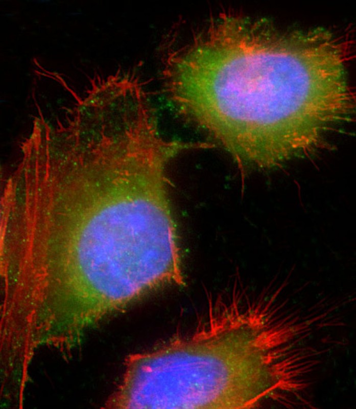 |
Immunofluorescent analysis of 4% paraformaldehyde-fixed, 0.1% Triton X-100 permeabilized HeLa (human cervical epithelial adenocarcinoma cell line) cells labeling HSD17B10 with P34365 at 1/25 dilution, followed by Dylight® 488-conjugated goat anti-rabbit IgG secondary antibody at 1/200 dilution (green). Immunofluorescence image showing cytoplasm and nucleus staining on HeLa cell line. Cytoplasmic actin is detected with Dylight® 554 Phalloidin at 1/100 dilution (red). The nuclear counter stain is DAPI (blue). |
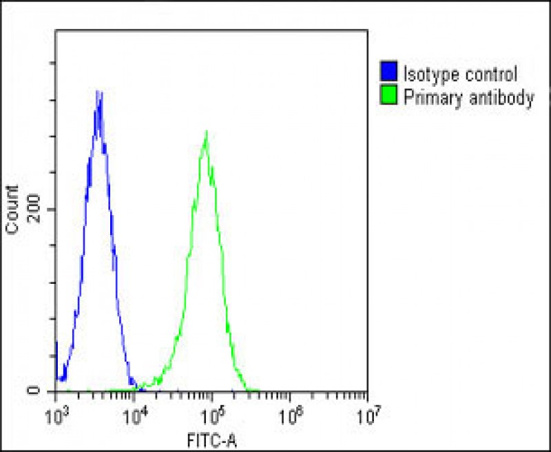 |
Overlay histogram showing Hela cells stained with P34365(green line). The cells were fixed with 2% paraformaldehyde (10 min) and then permeabilized with 90% methanol for 10 min. The cells were then icubated in 2% bovine serum albumin to block non-specific protein-protein interactions followed by the antibody (P34365, 1:25 dilution) for 60 min at 37ºC. The secondary antibody used was Goat-Anti-Rabbit IgG, DyLight® 488 Conjugated Highly Cross-Adsorbed(OH191631) at 1/200 dilution for 40 min at 37ºC. Isotype control antibody (blue line) was rabbit IgG1 (1μg/1×10^6 cells) used under the same conditions. Acquisition of >10, 000 events was performed. |
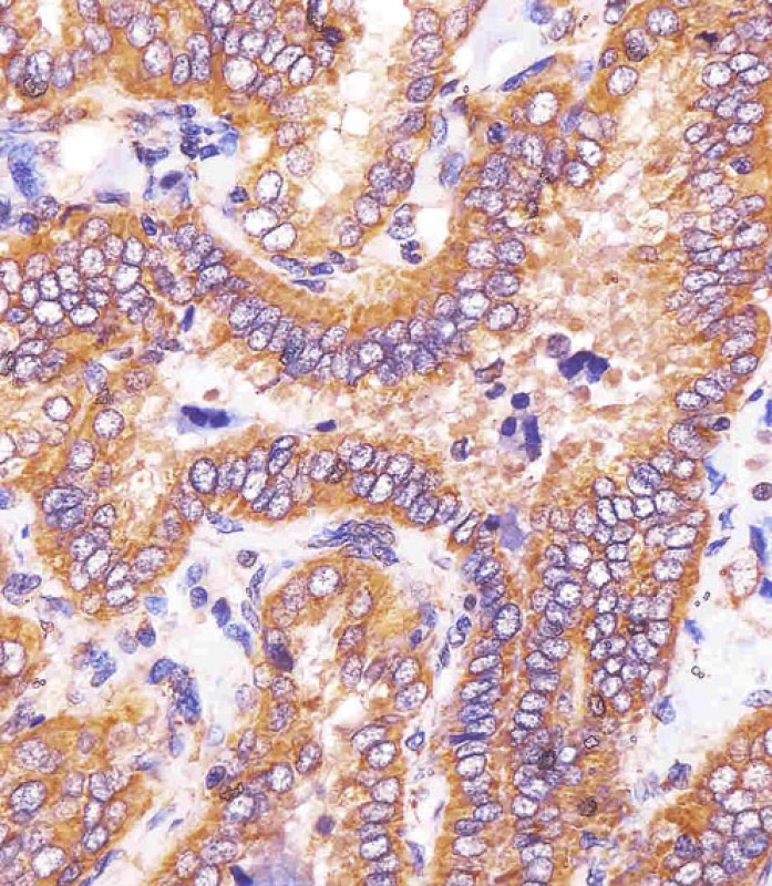 |
P34365 staining HSD17B10 in human thyroid carcinoma sections by Immunohistochemistry (IHC-P – paraformaldehyde-fixed, paraffin-embedded sections). Tissue was fixed with formaldehyde and blocked with 3% BSA for 0. 5 hour at room temperature; antigen retrieval was by heat mediation with a citrate buffer (pH6). Samples were incubated with primary antibody (1/25) for 1 hours at 37°C. A undiluted biotinylated goat polyvalent antibody was used as the secondary antibody. |
本公司的所有产品仅用于科学研究或者工业应用等非医疗目的,不可用于人类或动物的临床诊断或治疗,非药用,非食用。
暂无评论
本公司的所有产品仅用于科学研究或者工业应用等非医疗目的,不可用于人类或动物的临床诊断或治疗,非药用,非食用。


发表回复