 中文
中文 别名:Insulin-like growth factor 2 mRNA-binding protein 1, IGF2 mRNA-binding protein 1, IMP-1, IMP1, Coding region determinant-binding protein, CRD-BP, IGF-II mRNA-binding protein 1, VICKZ family member 1, Zipcode-binding protein 1, ZBP-1, IGF2BP1, CRDBP, VICKZ1, ZBP1应用:WB,FCM
反应种属:Human, Mouse, Rat
规格:50μl/100μl
| Description |
|---|
| IGF2BP1 is a member of the insulin-like growth factor 2 mRNA-binding protein family. The protein encoded by this gene contains four K homology domains and two RNA recognition motifs. It functions by binding to the mRNAs of certain genes, including insulin-like growth factor 2, beta-actin and beta-transducin repeat-containing protein, and regulating their translation. Two transcript variants encoding different isoforms have been found for this gene. |
| Specification | |
|---|---|
| Aliases | Insulin-like growth factor 2 mRNA-binding protein 1, IGF2 mRNA-binding protein 1, IMP-1, IMP1, Coding region determinant-binding protein, CRD-BP, IGF-II mRNA-binding protein 1, VICKZ family member 1, Zipcode-binding protein 1, ZBP-1, IGF2BP1, CRDBP, VICKZ1, ZBP1 |
| Entrez GeneID | 10642 |
| Swissprot | Q9NZI8 |
| WB Predicted band size | 63.5kDa |
| Host/Isotype | Rabbit IgG |
| Storage | Store at 4°C short term. Aliquot and store at -20°C long term. Avoid freeze/thaw cycles. |
| Species Reactivity | Human, Mouse, Rat |
| Immunogen | This IGF2BP1 antibody is generated from rabbits immunized with a KLH conjugated synthetic peptide between 508-534 amino acids from the C-terminal region of human IGF2BP1. |
| Formulation | Purified polyclonal antibody supplied in PBS with 0.05% sodium azide. This antibody is purified through a protein A column, followed by peptide affinity purification. |
| Application | |
|---|---|
| WB | 1/1000-1/2000 |
| FCM | 1/25 |
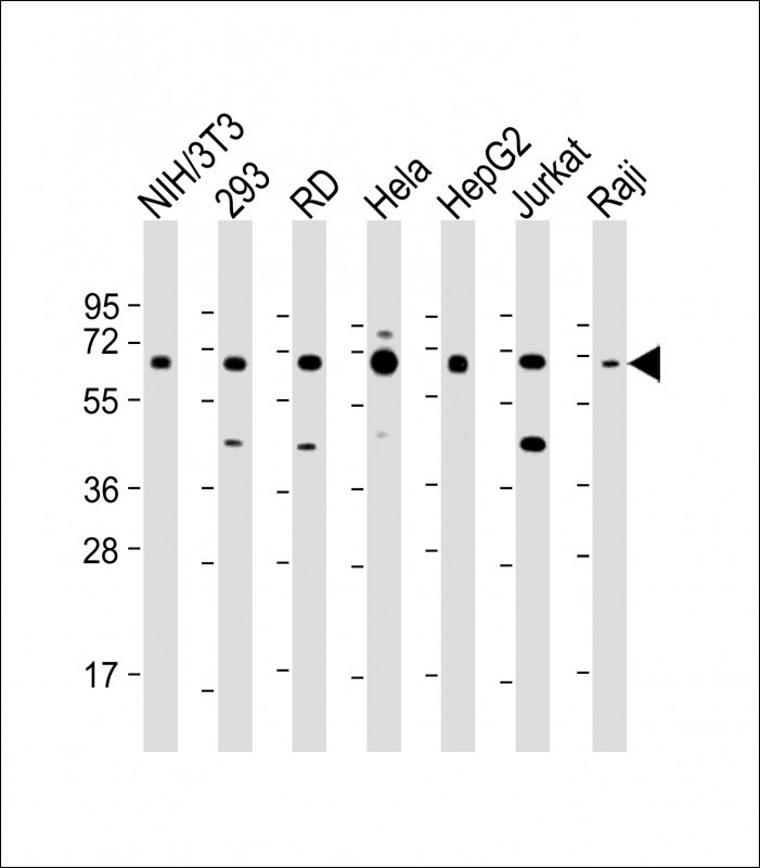 |
All lanes : Anti-IGF2BP1 Antibody (C-term) at 1:1000-1:2000 dilution Lane 1: NIH/3T3 whole cell lysate Lane 2: 293 whole cell lysate Lane 3: RD whole cell lysate Lane 4: Hela whole cell lysate Lane 5: HepG2 whole cell lysate Lane 6: Jurkat whole cell lysate Lane 7: Raji whole cell lysate Lysates/proteins at 20 µg per lane. Secondary Predicted band size : 63 kDa Blocking/Dilution buffer: 5% NFDM/TBST. |
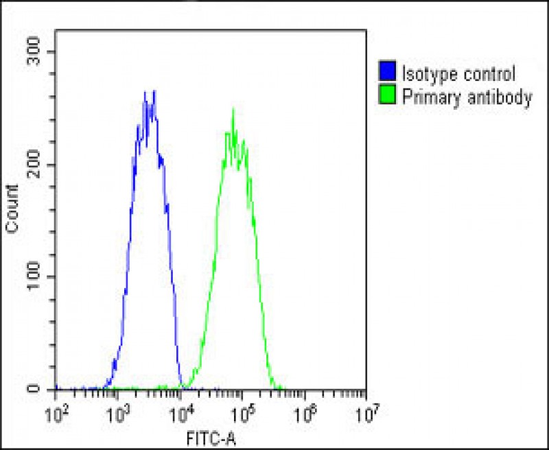 |
Overlay histogram showing HepG2 cells stained with P34349(green line). The cells were fixed with 2% paraformaldehyde (10 min) and then permeabilized with 90% methanol for 10 min. The cells were then icubated in 2% bovine serum albumin to block non-specific protein-protein interactions followed by the antibody (P34349, 1:25 dilution) for 60 min at 37ºC. The secondary antibody used was Goat-Anti-Rabbit IgG, DyLight® 488 Conjugated Highly Cross-Adsorbed(OH191631) at 1/200 dilution for 40 min at 37ºC. Isotype control antibody (blue line) was rabbit IgG1 (1μg/1×10^6 cells) used under the same conditions. Acquisition of >10, 000 events was performed. |
本公司的所有产品仅用于科学研究或者工业应用等非医疗目的,不可用于人类或动物的临床诊断或治疗,非药用,非食用。
暂无评论
本公司的所有产品仅用于科学研究或者工业应用等非医疗目的,不可用于人类或动物的临床诊断或治疗,非药用,非食用。


发表回复