 中文
中文 别名:Interleukin-12 receptor subunit beta-2, IL-12 receptor subunit beta-2, IL-12R subunit beta-2, IL-12R-beta-2, IL-12RB2, IL12RB2应用:WB,FCM
反应种属:Human, Mouse, Rat
规格:50μl/100μl
| Description |
|---|
| The protein encoded by this gene is a type I transmembrane protein identified as a subunit of the interleukin 12 receptor complex. The coexpression of this and IL12RB1 proteins was shown to lead to the formation of high-affinity IL12 binding sites and reconstitution of IL12 dependent signaling. The expression of this gene is up-regulated by interferon gamma in Th1 cells, and plays a role in Th1 cell differentiation. The up-regulation of this gene is found to be associated with a number of infectious diseases, such as Crohn’s disease and leprosy, which is thought to contribute to the inflammatory response and host defense. |
| Specification | |
|---|---|
| Aliases | Interleukin-12 receptor subunit beta-2, IL-12 receptor subunit beta-2, IL-12R subunit beta-2, IL-12R-beta-2, IL-12RB2, IL12RB2 |
| Entrez GeneID | 3595 |
| Swissprot | Q99665 |
| WB Predicted band size | 97.1kDa |
| Host/Isotype | Rabbit IgG |
| Storage | Store at 4°C short term. Aliquot and store at -20°C long term. Avoid freeze/thaw cycles. |
| Species Reactivity | Human, Mouse, Rat |
| Immunogen | This IL12_2 antibody is generated from rabbits immunized with a KLH conjugated synthetic peptide between 756-783 amino acids from the C-terminal region of human IL12_2. |
| Formulation | Purified polyclonal antibody supplied in PBS with 0.05% sodium azide. This antibody is purified through a protein A column, followed by peptide affinity purification. |
| Application | |
|---|---|
| WB | 1/1000 |
| FCM | 1/10-1/50 |
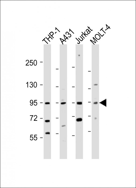 |
All lanes : Anti-IL12RB2 Antibody (C-term) at 1:2000 dilution Lane 1: THP-1 whole cell lysate Lane 2: A431 whole cell lysate Lane 3: Jurkat whole cell lysate Lane 4: MOLT-4 whole cell lysate Lysates/proteins at 20 µg per lane. Secondary Predicted band size : 97 kDa Blocking/Dilution buffer: 5% NFDM/TBST. |
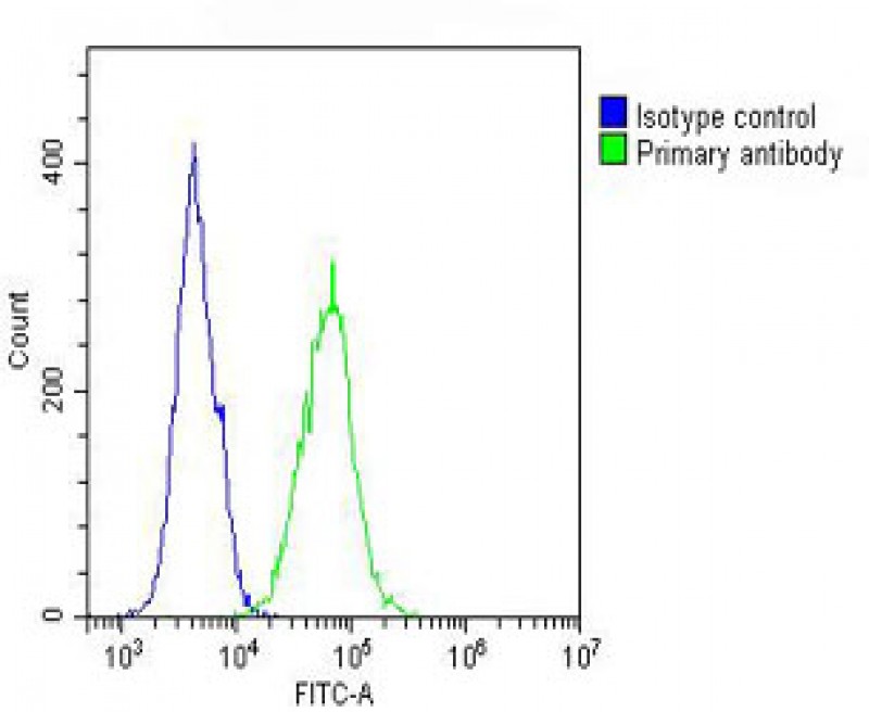 |
Overlay histogram showing A431 cells stained with P34310 (green line). The cells were fixed with 2% paraformaldehyde (10 min) and then permeabilized with 90% methanol for 10 min. The cells were then icubated in 2% bovine serum albumin to block non-specific protein-protein interactions followed by the antibody (P34310, 1:25 dilution) for 60 min at 37ºC. The secondary antibody used was Goat-Anti-Rabbit IgG, DyLight® 488 Conjugated Highly Cross-Adsorbed(OH191631) at 1/200 dilution for 40 min at 37ºC. Isotype control antibody (blue line) was rabbit IgG (1μg/1×10^6 cells) used under the same conditions. Acquisition of >10, 000 events was performed. |
本公司的所有产品仅用于科学研究或者工业应用等非医疗目的,不可用于人类或动物的临床诊断或治疗,非药用,非食用。
暂无评论
本公司的所有产品仅用于科学研究或者工业应用等非医疗目的,不可用于人类或动物的临床诊断或治疗,非药用,非食用。


发表回复