 中文
中文 别名:Interleukin-1 alpha, IL-1 alpha, Hematopoietin-1, IL1A, IL1F1应用:WB,IHC,FCM
反应种属:Human, Mouse
规格:50μl/100μl
| Description |
|---|
| IL1A is a member of the interleukin 1 cytokine family. This cytokine is a pleiotropic cytokine involved in various immune responses, inflammatory processes, and hematopoiesis. This cytokine is produced by monocytes and macrophages as a proprotein, which is proteolytically processed and released in response to cell injury, and thus induces apoptosis. |
| Specification | |
|---|---|
| Aliases | Interleukin-1 alpha, IL-1 alpha, Hematopoietin-1, IL1A, IL1F1 |
| Entrez GeneID | 3552 |
| Swissprot | P01583 |
| WB Predicted band size | 30.6kDa |
| Host/Isotype | Rabbit IgG |
| Storage | Store at 4°C short term. Aliquot and store at -20°C long term. Avoid freeze/thaw cycles. |
| Species Reactivity | Human, Mouse |
| Immunogen | This IL1A antibody is generated from rabbits immunized with a KLH conjugated synthetic peptide between 177-206 amino acids from the Central region of human IL1A. |
| Formulation | Purified polyclonal antibody supplied in PBS with 0.05% sodium azide. This antibody is purified through a protein A column, followed by peptide affinity purification. |
| Application | |
|---|---|
| WB | 1/1000 |
| IHC | 1/500 |
| FCM | 1/25 |
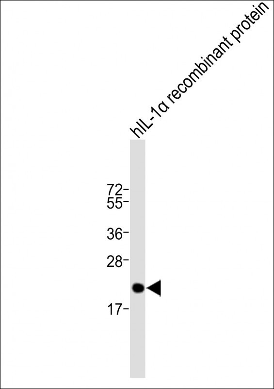 |
Anti-IL1A Antibody (Center) at 1:2000 dilution + hIL-1α recombinant protein
Lysates/proteins at 20 ng per lane. Secondary Predicted band size : 31 kDa Blocking/Dilution buffer: 5% NFDM/TBST. |
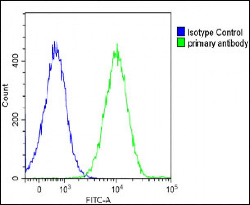 |
Overlay histogram showing Ramos cells stained with P34543(green line). The cells were fixed with 2% paraformaldehyde and then permeabilized with 90% methanol for 10 min. The cells were then icubated in 2% bovine serum albumin to block non-specific protein-protein interactions followed by the antibody (1:25 dilution) for 60 min at 37ºC. The secondary antibody used was Goat-Anti-Rabbit IgG, DyLight® 488 Conjugated Highly Cross-Adsorbed at 1/200 dilution for 40 min at Room temperature. Isotype control antibody (blue line) was rabbit IgG1 (1μg/1×10^6 cells) used under the same conditions. Acquisition of >10, 000 events was performed. |
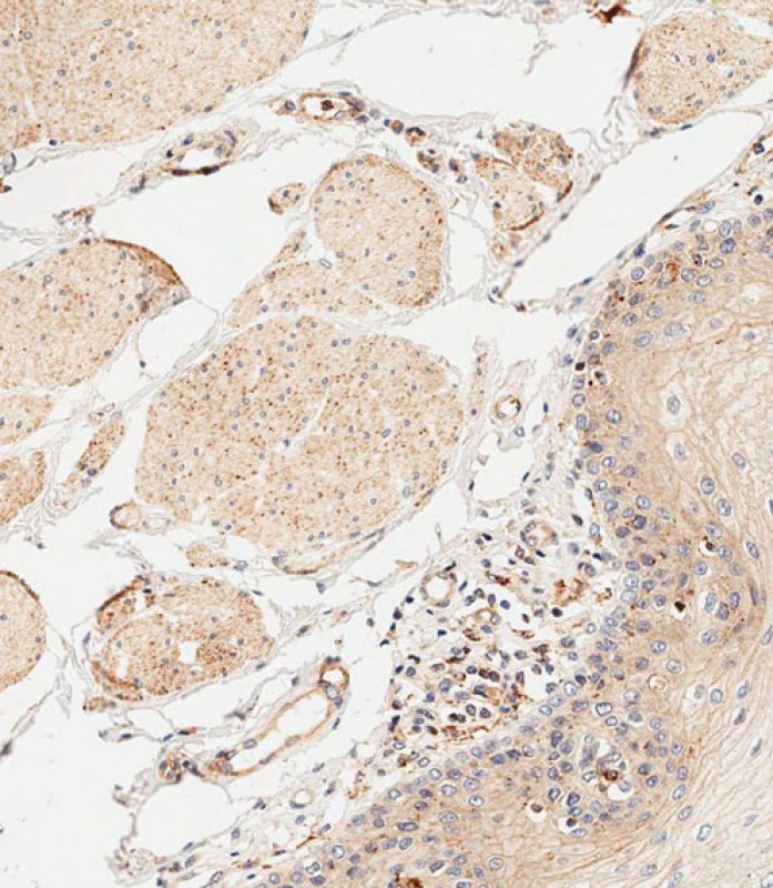 |
Immunohistochemical analysis of paraffin-embedded human esophagus tissue using P34543 performed on the Leica® BOND RXm. Samples were incubated with primary antibody(1/500) for 1 hours at room temperature. A undiluted biotinylated CRF Anti-Polyvalent HRP Polymer antibody was used as the secondary antibody. |
本公司的所有产品仅用于科学研究或者工业应用等非医疗目的,不可用于人类或动物的临床诊断或治疗,非药用,非食用。
暂无评论
本公司的所有产品仅用于科学研究或者工业应用等非医疗目的,不可用于人类或动物的临床诊断或治疗,非药用,非食用。


发表回复