 中文
中文 别名:Interleukin-1 receptor-like 2, IL-36 receptor, IL-36R, Interleukin-1 receptor-related protein 2, IL-1Rrp2, IL1R-rp2, IL1RL2, IL1RRP2应用:WB,FCM
反应种属:Human
规格:50μl/100μl
| Description |
|---|
| The protein encoded by this gene is a member of the interleukin 1 receptor family. An experiment with transient gene expression demonstrated that this receptor was incapable of binding to interleukin 1 alpha and interleukin 1 beta with high affinity. This gene and four other interleukin 1 receptor family genes, including interleukin 1 receptor, type I (IL1R1), interleukin 1 receptor, type II (IL1R2), interleukin 1 receptor-like 1 (IL1RL1), and interleukin 18 receptor 1 (IL18R1), form a cytokine receptor gene cluster in a region mapped to chromosome 2q12. [provided by RefSeq]. |
| Specification | |
|---|---|
| Aliases | Interleukin-1 receptor-like 2, IL-36 receptor, IL-36R, Interleukin-1 receptor-related protein 2, IL-1Rrp2, IL1R-rp2, IL1RL2, IL1RRP2 |
| Entrez GeneID | 8808 |
| Swissprot | Q9HB29 |
| WB Predicted band size | 65.4kDa |
| Host/Isotype | Rabbit IgG |
| Storage | Store at 4°C short term. Aliquot and store at -20°C long term. Avoid freeze/thaw cycles. |
| Species Reactivity | Human |
| Immunogen | This IL1RL2 antibody is generated from rabbits immunized with a KLH conjugated synthetic peptide between 257-286 amino acids from the Central region of human IL1RL2. |
| Formulation | Purified polyclonal antibody supplied in PBS with 0.05% sodium azide. This antibody is purified through a protein A column, followed by peptide affinity purification. |
| Application | |
|---|---|
| WB | 1/1000 |
| FCM | 1/25 |
 |
|
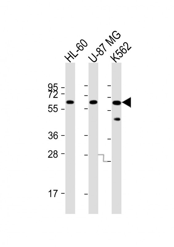 |
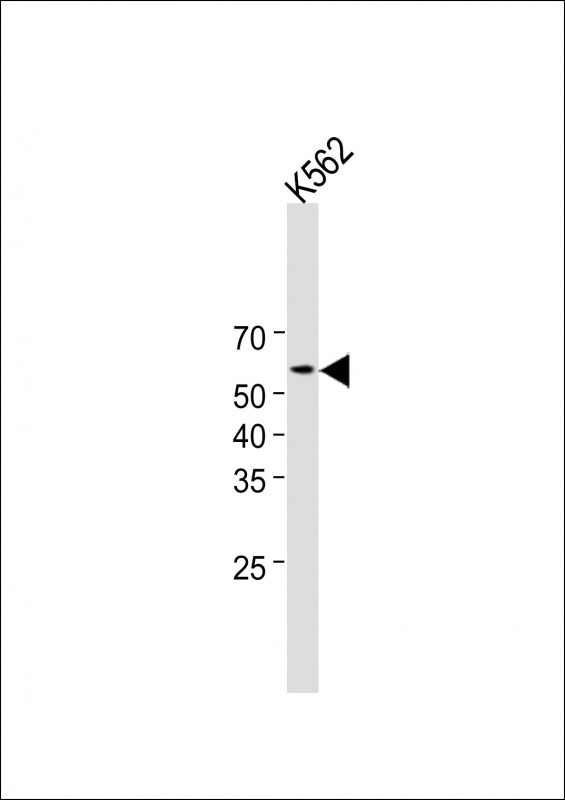
All lanes : Anti-IL1RL2 Antibody (Center) at 1:8000 dilution Lane 1: HL-60 whole cell lysate Lane 2: U-87 MG whole cell lysate Lane 3: K562 whole cell lysate Lysates/proteins at 20 µg per lane. Secondary Predicted band size : 65 kDa Blocking/Dilution buffer: 5% NFDM/TBST. |
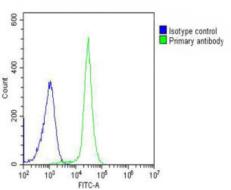 |
Overlay histogram showing K562 cells stained with P35095 (green line). The cells were fixed with 2% paraformaldehyde (10 min). The cells were then icubated in 2% bovine serum albumin to block non-specific protein-protein interactions followed by the antibody (P35095, 1:25 dilution) for 60 min at 37ºC. The secondary antibody used was Goat-Anti-Rabbit IgG, DyLight® 488 Conjugated Highly Cross-Adsorbed(OH191631) at 1/200 dilution for 40 min at 37ºC. Isotype control antibody (blue line) was rabbit IgG (1μg/1×10^6 cells) used under the same conditions. Acquisition of >10, 000 events was performed. |
本公司的所有产品仅用于科学研究或者工业应用等非医疗目的,不可用于人类或动物的临床诊断或治疗,非药用,非食用。
暂无评论
本公司的所有产品仅用于科学研究或者工业应用等非医疗目的,不可用于人类或动物的临床诊断或治疗,非药用,非食用。


发表回复