 中文
中文 别名:Inter-alpha-trypsin inhibitor heavy chain H4, ITI heavy chain H4, ITI-HC4, Inter-alpha-inhibitor heavy chain 4, Inter-alpha-trypsin inhibitor family heavy chain-related protein, IHRP, Plasma kallikrein sensitive glycoprotein 120, Gp120, PK-120, 70 kDa inter-alpha-trypsin inhibitor heavy chain H4, 35 kDa inter-alpha-trypsin inhibitor heavy chain H4, ITIH4, IHRP, ITIHL1, PK120应用:WB,ICC,FCM
反应种属:Human
规格:50μl/100μl
| Description |
|---|
| Type II acute-phase protein (APP) involved in inflammatory responses to trauma. May also play a role in liver development or regeneration. |
| Specification | |
|---|---|
| Aliases | Inter-alpha-trypsin inhibitor heavy chain H4, ITI heavy chain H4, ITI-HC4, Inter-alpha-inhibitor heavy chain 4, Inter-alpha-trypsin inhibitor family heavy chain-related protein, IHRP, Plasma kallikrein sensitive glycoprotein 120, Gp120, PK-120, 70 kDa inter-alpha-trypsin inhibitor heavy chain H4, 35 kDa inter-alpha-trypsin inhibitor heavy chain H4, ITIH4, IHRP, ITIHL1, PK120 |
| Entrez GeneID | 3700 |
| Swissprot | Q14624 |
| WB Predicted band size | 103.4kDa |
| Host/Isotype | Rabbit IgG |
| Storage | Store at 4°C short term. Aliquot and store at -20°C long term. Avoid freeze/thaw cycles. |
| Species Reactivity | Human |
| Immunogen | This ITIH4 antibody is generated from a rabbit immunized with a KLH conjugated synthetic peptide between 784-816 amino acids from human ITIH4. |
| Application | |
|---|---|
| WB | 1/2000 |
| ICC | 1/25 |
| FCM | 1/25 |
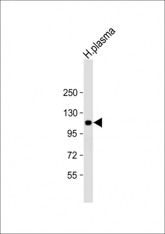 |
Anti-ITIH4 Antibody (C-Term) at 1:2000 dilution + human plasma lysate
Lysates/proteins at 20 µg per lane. Secondary Predicted band size : 103 kDa Blocking/Dilution buffer: 5% NFDM/TBST. |
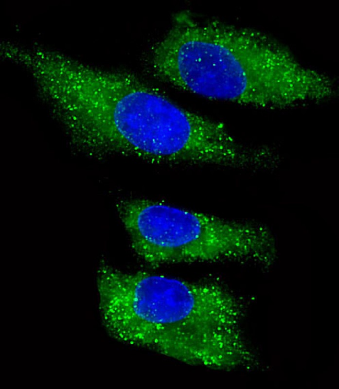 |
Immunofluorescent analysis of 4% paraformaldehyde-fixed, 0.1% Triton X-100 permeabilized HepG2 (human liver hepatocellular carcinoma cell line) cells labeling ITIH4 with P33949 at 1/25 dilution, followed by Dylight® 488-conjugated goat anti-rabbit IgG secondary antibody at 1/200 dilution (green). Immunofluorescence image showing cytoplasm staining on HepG2 cell line. The nuclear counter stain is DAPI (blue). |
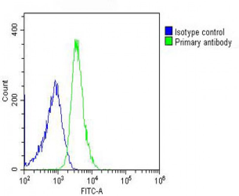 |
Overlay histogram showing U-2OS cells stained with P33949 (green line). The cells were fixed with 2% paraformaldehyde (10 min) and then permeabilized with 90% methanol for 10 min. The cells were then icubated in 2% bovine serum albumin to block non-specific protein-protein interactions followed by the antibody (P33949, 1:25 dilution) for 60 min at 37ºC. The secondary antibody used was Goat-Anti-Rabbit IgG, DyLight® 488 Conjugated Highly Cross-Adsorbed(OH191631) at 1/200 dilution for 40 min at 37ºC. Isotype control antibody (blue line) was rabbit IgG (1μg/1×10^6 cells) used under the same conditions. Acquisition of >10, 000 events was performed. |
本公司的所有产品仅用于科学研究或者工业应用等非医疗目的,不可用于人类或动物的临床诊断或治疗,非药用,非食用。
暂无评论
本公司的所有产品仅用于科学研究或者工业应用等非医疗目的,不可用于人类或动物的临床诊断或治疗,非药用,非食用。


发表回复