 中文
中文 别名:BTB/POZ domain-containing protein KCTD11, KCTD11, C17orf36, REN应用:WB,IHC
反应种属:Human, Mouse
规格:50μl/100μl
| Description |
|---|
| Plays a role as a marker and a regulator of neuronal differentiation; Up-regulated by a variety of neurogenic signals, such as retinoic acid, epidermal growth factor/EGF and NGFB/nerve growth factor. Induces apoptosis, growth arrest and the expression of cyclin-dependent kinase inhibitor CDKN1B. Plays a role as a tumor repressor and inhibits cell growth and tumorigenicity of medulloblastoma (MDB). Acts as an E3 ubiquitin-protein ligase towards HDAC1, leading to its proteasomal degradation. Functions as antagonist of the Hedgehog pathway on cell proliferation and differentiation by affecting the nuclear transfer of transcrition factor GLI1, thus maintaining cerebellar granule cells in undifferentiated state, this effect probably occurs via HDAC1 down-regulation, keeping GLI1 acetylated and inactive. When knock- down, Hedgehog antagonism is impaired and proliferation of granule cells is sustained. Activates the caspase cascade. |
| Specification | |
|---|---|
| Aliases | BTB/POZ domain-containing protein KCTD11, KCTD11, C17orf36, REN |
| Entrez GeneID | 147040 |
| Swissprot | Q693B1 |
| WB Predicted band size | 25.9kDa |
| Host/Isotype | Rabbit IgG |
| Storage | Store at 4°C short term. Aliquot and store at -20°C long term. Avoid freeze/thaw cycles. |
| Species Reactivity | Human, Mouse |
| Immunogen | This KCTD11 antibody is generated from a rabbit immunized with a KLH conjugated synthetic peptide between 21-53 amino acids from human KCTD11. |
| Application | |
|---|---|
| WB | 1/1000-1/2000 |
| IHC | 1/100-1/500 |
 |
All lanes : Anti-KCTD11 Antibody (N-Term) at 1:1000-1:2000 dilution Lane 1: human brain lysate Lane 2: human cerebellum lysate Lane 3: mouse cerebellum lysate Lysates/proteins at 20 µg per lane. Secondary Predicted band size : 26 kDa Blocking/Dilution buffer: 5% NFDM/TBST. |
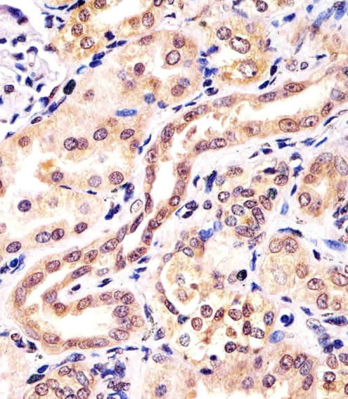 |
P33549 staining KCTD11 in human kidney tissue sections by Immunohistochemistry (IHC-P – paraformaldehyde-fixed, paraffin-embedded sections). Tissue was fixed with formaldehyde and blocked with 3% BSA for 0. 5 hour at room temperature; antigen retrieval was by heat mediation with a citrate buffer (pH6). Samples were incubated with primary antibody (1/25) for 1 hours at 37°C. A undiluted biotinylated goat polyvalent antibody was used as the secondary antibody. |
本公司的所有产品仅用于科学研究或者工业应用等非医疗目的,不可用于人类或动物的临床诊断或治疗,非药用,非食用。
暂无评论
本公司的所有产品仅用于科学研究或者工业应用等非医疗目的,不可用于人类或动物的临床诊断或治疗,非药用,非食用。


发表回复