 中文
中文 别名:Keratin, type II cytoskeletal 1, 67 kDa cytokeratin, Cytokeratin-1, CK-1, Hair alpha protein, Keratin-1, K1, Type-II keratin Kb1, KRT1, KRTA应用:WB,IHC,FCM
反应种属:Human
规格:50μl/100μl
| Description |
|---|
| KRT1 is a member of the keratin gene family. The type II cytokeratins consist of basic or neutral proteins which are arranged in pairs of heterotypic keratin chains coexpressed during differentiation of simple and stratified epithelial tissues. This type II cytokeratin is specifically expressed in the spinous and granular layers of the epidermis with family member KRT10 and mutations in these genes have been associated with bullous congenital ichthyosiform erythroderma. |
| Specification | |
|---|---|
| Aliases | Keratin, type II cytoskeletal 1, 67 kDa cytokeratin, Cytokeratin-1, CK-1, Hair alpha protein, Keratin-1, K1, Type-II keratin Kb1, KRT1, KRTA |
| Entrez GeneID | 3848 |
| Swissprot | P04264 |
| WB Predicted band size | 66.0kDa |
| Host/Isotype | Rabbit IgG |
| Storage | Store at 4°C short term. Aliquot and store at -20°C long term. Avoid freeze/thaw cycles. |
| Species Reactivity | Human |
| Immunogen | This KRT1 antibody is generated from rabbits immunized with a KLH conjugated synthetic peptide between 415-443 amino acids from the Central region of human KRT1. |
| Formulation | Purified polyclonal antibody supplied in PBS with 0.05% sodium azide. This antibody is prepared by Saturated Ammonium Sulfate (SAS) precipitation followed by dialysis against PBS. |
| Application | |
|---|---|
| WB | 1/1000 |
| IHC | 1/100-1/500 |
| FCM | 1/10-1/50 |
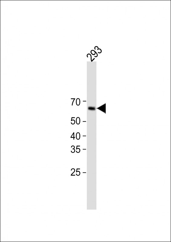 |
All lanes: Anti-KRT1 Antibody (Center) at 1:1000 dilution + 293 whole cell lysate
Lysates/proteins at 20 µg per lane. Observed band size: 66KDa Blocking/Dilution buffer: 5% NFDM/TBST. |
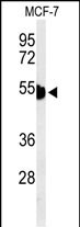 |
Western blot analysis of KRT1 Antibody (Center) (Cat. #P35092) in MCF-7 cell line lysates (35ug/lane). KRT1 (arrow) was detected using the purified Pab. |
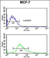 |
KRT1 Antibody (Center) (Cat. #P35092) flow cytometric analysis of MCF-7 cells (bottom histogram) compared to a negative control cell (top histogram).FITC-conjugated goat-anti-rabbit secondary antibodies were used for the analysis. |
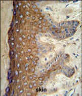 |
KRT1 Antibody (Center) (Cat. #P35092) IHC analysis in formalin fixed and paraffin embedded skin tissue followed by peroxidase conjugation of the secondary antibody and DAB staining. This data demonstrates the use of the KRT1 Antibody (Center) for immunohistochemistry. Clinical relevance has not been evaluated. |
本公司的所有产品仅用于科学研究或者工业应用等非医疗目的,不可用于人类或动物的临床诊断或治疗,非药用,非食用。
暂无评论
本公司的所有产品仅用于科学研究或者工业应用等非医疗目的,不可用于人类或动物的临床诊断或治疗,非药用,非食用。


发表回复