 中文
中文 别名:Microtubule-associated proteins 1A/1B light chain 3A, Autophagy-related protein LC3 A, Autophagy-related ubiquitin-like modifier LC3 A, MAP1 light chain 3-like protein 1, MAP1A/MAP1B light chain 3 A, MAP1A/MAP1B LC3 A, Microtubule-associated protein 1 light chain 3 alpha, MAP1LC3A应用:WB,IHC
反应种属:Human, Mouse, Rat
规格:50μl/100μl
| Description |
|---|
| Ubiquitin-like modifier involved in formation of autophagosomal vacuoles (autophagosomes). Whereas LC3s are involved in elongation of the phagophore membrane, the GABARAP/GATE-16 subfamily is essential for a later stage in autophagosome maturation. |
| Specification | |
|---|---|
| Aliases | Microtubule-associated proteins 1A/1B light chain 3A, Autophagy-related protein LC3 A, Autophagy-related ubiquitin-like modifier LC3 A, MAP1 light chain 3-like protein 1, MAP1A/MAP1B light chain 3 A, MAP1A/MAP1B LC3 A, Microtubule-associated protein 1 light chain 3 alpha, MAP1LC3A |
| Entrez GeneID | 84557 |
| Swissprot | Q9H492 |
| WB Predicted band size | 14.3kDa |
| Host/Isotype | Rabbit IgG |
| Storage | Store at 4°C short term. Aliquot and store at -20°C long term. Avoid freeze/thaw cycles. |
| Species Reactivity | Human, Mouse, Rat |
| Immunogen | This antibody is generated from a rabbit immunized with a KLH conjugated synthetic peptide between 1-40 amino acids from human. |
| Application | |
|---|---|
| WB | 1/2000 |
| IHC | 1/500 |
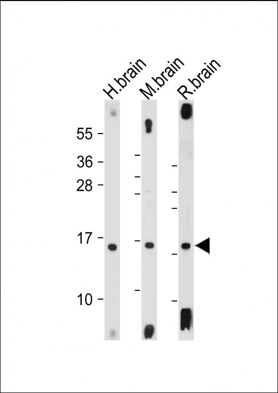 |
All lanes : Anti-MAP1LC3A antibody(N-term) at 1:2000 dilution Lane 1: Human brain tissue lysate Lane 2: Mouse brain tissue lysate Lane 3: Rat brain tissue lysate Lysates/proteins at 20 µg per lane. Secondary Predicted band size : 14 kDa Blocking/Dilution buffer: 5% NFDM/TBST. |
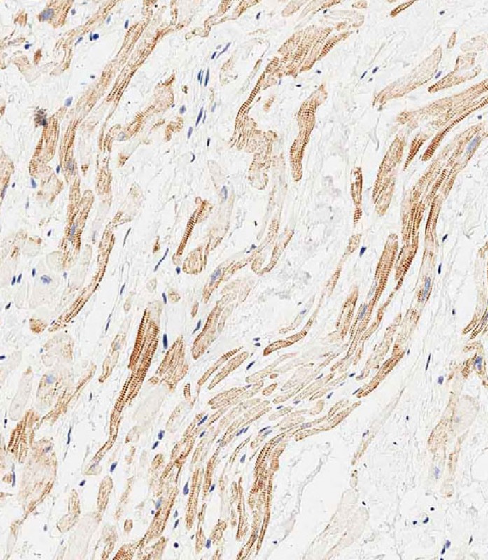 |
Immunohistochemical analysis of paraffin-embedded human heart tissue using P34668 performed on the Leica® BOND RXm. Tissue was fixed with formaldehyde at room temperature, antigen retrieval was by heat mediation with a EDTA buffer (pH9. 0). Samples were incubated with primary antibody(1:500) for 1 hours at room temperature. A undiluted biotinylated CRF Anti-Polyvalent HRP Polymer antibody was used as the secondary antibody. |
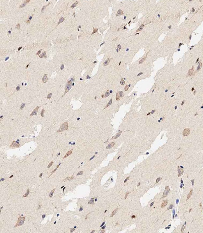 |
Immunohistochemical analysis of paraffin-embedded human brain tissue using P34668 performed on the Leica® BOND RXm. Tissue was fixed with formaldehyde at room temperature, antigen retrieval was by heat mediation with a EDTA buffer (pH9. 0). Samples were incubated with primary antibody(1:500) for 1 hours at room temperature. A undiluted biotinylated CRF Anti-Polyvalent HRP Polymer antibody was used as the secondary antibody. |
本公司的所有产品仅用于科学研究或者工业应用等非医疗目的,不可用于人类或动物的临床诊断或治疗,非药用,非食用。
暂无评论
本公司的所有产品仅用于科学研究或者工业应用等非医疗目的,不可用于人类或动物的临床诊断或治疗,非药用,非食用。


发表回复