 中文
中文 别名:Induced myeloid leukemia cell differentiation protein Mcl-1, Bcl-2-like protein 3, Bcl2-L-3, Bcl-2-related protein EAT/mcl1, mcl1/EAT, MCL1, BCL2L3应用:WB,IHC,FCM
反应种属:Human, Mouse, Rat
规格:50μl/100μl
| Description |
|---|
| The Mcl-1 protein belongs to the Bcl-2 family. Alternative splicing occurs at this locus and two transcript variants encoding distinct isoforms have been identified. The longer gene product (isoform 1) enhances cell survival by inhibiting apoptosis while the alternatively spliced shorter gene product (isoform 2) promotes apoptosis and is death-inducing. |
| Specification | |
|---|---|
| Aliases | Induced myeloid leukemia cell differentiation protein Mcl-1, Bcl-2-like protein 3, Bcl2-L-3, Bcl-2-related protein EAT/mcl1, mcl1/EAT, MCL1, BCL2L3 |
| Entrez GeneID | 4170 |
| Swissprot | Q07820 |
| WB Predicted band size | 37.3kDa |
| Host/Isotype | Rabbit IgG |
| Storage | Store at 4°C short term. Aliquot and store at -20°C long term. Avoid freeze/thaw cycles. |
| Species Reactivity | Human, Mouse, Rat |
| Immunogen | This MCL1 antibody is generated from rabbits immunized with a KLH conjugated synthetic peptide between 191-226 amino acids from human MCL1. |
| Formulation | Purified polyclonal antibody supplied in PBS with 0.05% sodium azide. This antibody is purified through a protein A column, followed by peptide affinity purification. |
| Application | |
|---|---|
| WB | 1/1000 |
| IHC | 1/100-1/500 |
| FCM | 1/25 |
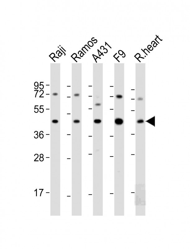 |
All lanes : Anti-MCL1 Antibody (BH3 Domain Specific) at 1:2000 dilution Lane 1: Raji whole cell lysate Lane 2: Ramos whole cell lysate Lane 3: A431 whole cell lysate Lane 4: F9 whole cell lysate Lane 5: rat heart lysate Lysates/proteins at 20 µg per lane. Secondary Predicted band size : 37 kDa Blocking/Dilution buffer: 5% NFDM/TBST. |
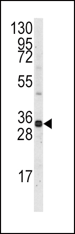 |
Western blot analysis of anti-Mcl-1 BH3 Domain Pab (Cat. #P34313) in Ramos cell line lysates (35ug/lane). Mcl-1-BH3(arrow) was detected using the purified Pab. |
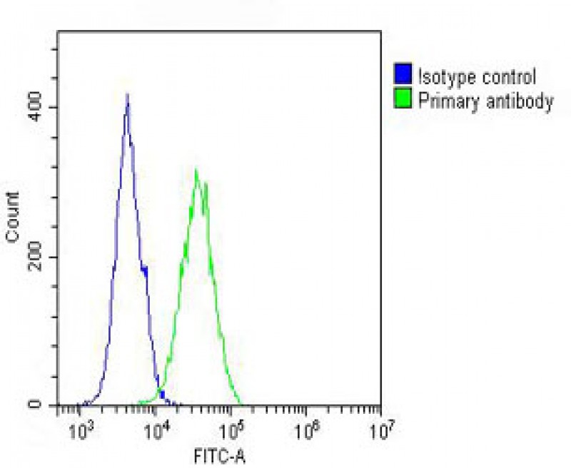 |
Overlay histogram showing A431 cells stained with P34313 (green line). The cells were fixed with 2% paraformaldehyde (10 min) and then permeabilized with 90% methanol for 10 min. The cells were then icubated in 2% bovine serum albumin to block non-specific protein-protein interactions followed by the antibody (P34313, 1:25 dilution) for 60 min at 37ºC. The secondary antibody used was Goat-Anti-Rabbit IgG, DyLight® 488 Conjugated Highly Cross-Adsorbed(OH191631) at 1/200 dilution for 40 min at 37ºC. Isotype control antibody (blue line) was rabbit IgG (1μg/1×10^6 cells) used under the same conditions. Acquisition of >10, 000 events was performed. |
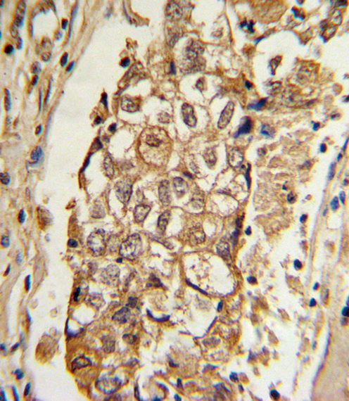 |
Formalin-fixed and paraffin-embedded human breast carcinoma with MCL1 Antibody (BH3 Domain Specific), which was peroxidase-conjugated to the secondary antibody, followed by DAB staining. This data demonstrates the use of this antibody for immunohistochemistry; clinical relevance has not been evaluated. |
本公司的所有产品仅用于科学研究或者工业应用等非医疗目的,不可用于人类或动物的临床诊断或治疗,非药用,非食用。
暂无评论
本公司的所有产品仅用于科学研究或者工业应用等非医疗目的,不可用于人类或动物的临床诊断或治疗,非药用,非食用。


发表回复