 中文
中文 别名:Meckel syndrome type 1 protein, MKS1应用:WB,ICC,FCM
反应种属:Human
规格:50μl/100μl
| Description |
|---|
| Component of the tectonic-like complex, a complex localized at the transition zone of primary cilia and acting as a barrier that prevents diffusion of transmembrane proteins between the cilia and plasma membranes. Involved in centrosome migration to the apical cell surface during early ciliogenesis. Required for ciliary structure and function, including a role in regulating length and appropriate number through modulating centrosome duplication. Required for cell branching morphology. |
| Specification | |
|---|---|
| Aliases | Meckel syndrome type 1 protein, MKS1 |
| Entrez GeneID | 54903 |
| Swissprot | Q9NXB0 |
| WB Predicted band size | 64.5kDa |
| Host/Isotype | Rabbit IgG |
| Storage | Store at 4°C short term. Aliquot and store at -20°C long term. Avoid freeze/thaw cycles. |
| Species Reactivity | Human |
| Immunogen | This MKS1 antibody is generated from a rabbit immunized with a KLH conjugated synthetic peptide between 90-124 amino acids from the human region of human MKS1. |
| Application | |
|---|---|
| WB | 1/2000 |
| ICC | 1/25 |
| FCM | 1/25 |
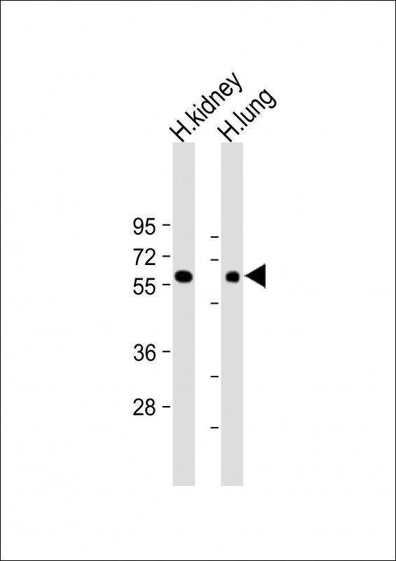 |
All lanes : Anti-MKS1 Antibody (N-Term) at 1:2000 dilution Lane 1: Human kidney lysate Lane 2: Human lung lysate Lysates/proteins at 20 µg per lane. Secondary Predicted band size : 65 kDa Blocking/Dilution buffer: 5% NFDM/TBST. |
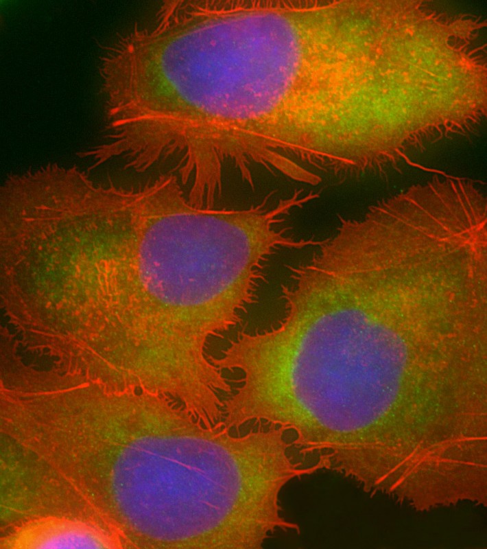 |
Immunofluorescent analysis of 4% paraformaldehyde-fixed, 0.1% Triton X-100 permeabilized HepG2 (human liver hepatocellular carcinoma cell line) cells labeling MKS1 with P34515 at 1/25 dilution, followed by Dylight® 488-conjugated goat anti-rabbit IgG secondary antibody at 1/200 dilution (green). Immunofluorescence image showing cytoplasm staining on HepG2 cell line. Cytoplasmic actin is detected with Dylight® 554 Phalloidin at 1/100 dilution (red). The nuclear counter stain is DAPI (blue). |
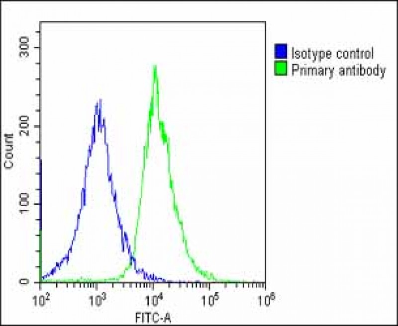 |
Overlay histogram showing HepG2 cells stained with P34515(green line). The cells were fixed with 2% paraformaldehyde (10 min) and then permeabilized with 90% methanol for 10 min. The cells were then icubated in 2% bovine serum albumin to block non-specific protein-protein interactions followed by the antibody (P34515, 1:25 dilution) for 60 min at 37ºC. The secondary antibody used was Goat-Anti-Rabbit IgG, DyLight® 488 Conjugated Highly Cross-Adsorbed(OE188374) at 1/200 dilution for 40 min at 37ºC. Isotype control antibody (blue line) was rabbit IgG1 (1μg/1×10^6 cells) used under the same conditions. Acquisition of >10, 000 events was performed. |
本公司的所有产品仅用于科学研究或者工业应用等非医疗目的,不可用于人类或动物的临床诊断或治疗,非药用,非食用。
暂无评论
本公司的所有产品仅用于科学研究或者工业应用等非医疗目的,不可用于人类或动物的临床诊断或治疗,非药用,非食用。


发表回复