 中文
中文 别名:Macrophage colony-stimulating factor 1 receptor, CSF-1 receptor, CSF-1-R, CSF-1R, M-CSF-R, Proto-oncogene c-Fms, CD115, Csf1r, Csfmr, Fms应用:WB,ICC
反应种属:Mouse, Rat
规格:50μl/100μl
| Description |
|---|
| Csf1r is a protein tyrosine-kinase transmembrane receptor for CSF1 and IL34. |
| Specification | |
|---|---|
| Aliases | Macrophage colony-stimulating factor 1 receptor, CSF-1 receptor, CSF-1-R, CSF-1R, M-CSF-R, Proto-oncogene c-Fms, CD115, Csf1r, Csfmr, Fms |
| Entrez GeneID | 12978 |
| Swissprot | P09581 |
| WB Predicted band size | 109.2kDa |
| Host/Isotype | Rabbit IgG |
| Storage | Store at 4°C short term. Aliquot and store at -20°C long term. Avoid freeze/thaw cycles. |
| Species Reactivity | Mouse, Rat |
| Immunogen | This Mouse Csf1r antibody is generated from rabbits immunized with a KLH conjugated synthetic peptide between 895-923 amino acids from the C-terminal region of mouse Csf1r. |
| Formulation | Purified polyclonal antibody supplied in PBS with 0.05% sodium azide. This antibody is purified through a protein A column, followed by peptide affinity purification. |
| Application | |
|---|---|
| WB | 1/2000 |
| ICC | 1/25 |
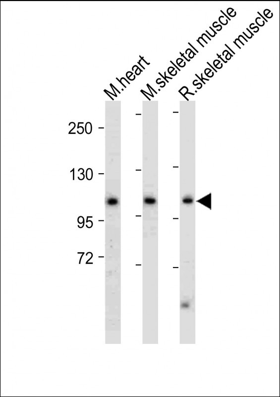 |
All lanes : Anti-Mouse Csf1r Antibody (C-term) at 1:2000 dilution Lane 1: Mouse heart tissue lysate Lane 2: Mouse skeletal muscle tissue lysate Lane 3: Rat skeletal muscle tissue lysate Lysates/proteins at 20 µg per lane. Secondary Predicted band size : 109 kDa Blocking/Dilution buffer: 5% NFDM/TBST. |
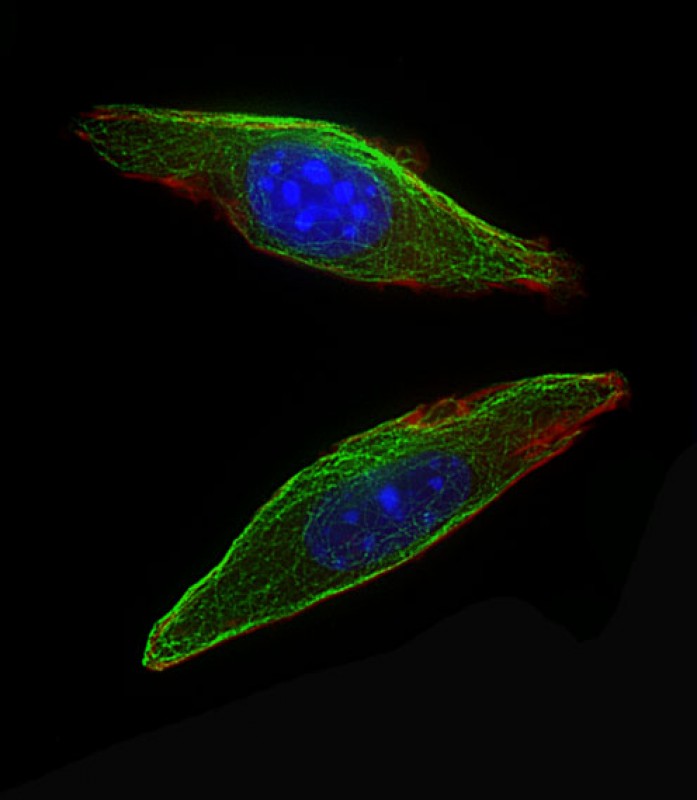 |
Immunofluorescent analysis of 4% paraformaldehyde-fixed, 0. 1% Triton X-100 permeabilized NIH/3T3 cells labeling Csf1r with P34679 at 1/25 dilution, followed by Dylight® 488-conjugated goat anti-Rabbit IgG secondary antibody at 1/200 dilution (green). Immunofluorescence image showing Cytoplasm staining on NIH/3T3 cell line. Cytoplasmic actin is detected with Dylight® 554 Phalloidin(red). The nuclear counter stain is DAPI (blue). |
本公司的所有产品仅用于科学研究或者工业应用等非医疗目的,不可用于人类或动物的临床诊断或治疗,非药用,非食用。
暂无评论
本公司的所有产品仅用于科学研究或者工业应用等非医疗目的,不可用于人类或动物的临床诊断或治疗,非药用,非食用。


发表回复