 中文
中文 别名:Homeobox protein Nkx-25, Cardiac-specific homeobox, Homeobox protein CSX, Homeobox protein NK-2 homolog E, Nkx2-5, Csx, Nkx-25, Nkx2e应用:WB,IHC,FCM
反应种属:Human, Mouse, Rat
规格:50μl/100μl
| Description |
|---|
| Implicated in commitment to and/or differentiation of the myocardial lineage. Acts as a transcriptional activator of ANF in cooperation with GATA4. It is transcriptionally controlled by PBX1 and acts as a transcriptional repressor of CDKN2B. Together with PBX1, it is required for spleen development through a mechanism that involves CDKN2B repression. |
| Specification | |
|---|---|
| Aliases | Homeobox protein Nkx-25, Cardiac-specific homeobox, Homeobox protein CSX, Homeobox protein NK-2 homolog E, Nkx2-5, Csx, Nkx-25, Nkx2e |
| Entrez GeneID | 18091 |
| Swissprot | P42582 |
| WB Predicted band size | 34.2kDa |
| Host/Isotype | Rabbit IgG |
| Storage | Store at 4°C short term. Aliquot and store at -20°C long term. Avoid freeze/thaw cycles. |
| Species Reactivity | Human, Mouse, Rat |
| Immunogen | This mouse Nkx2-5 antibody is generated from a rabbit immunized with a KLH conjugated synthetic peptide between 98-133 amino acids from the Central region of mouse Nkx2-5. |
| Formulation | Purified polyclonal antibody supplied in PBS with 0.05% sodium azide. This antibody is purified through a protein A column, followed by peptide affinity purification. |
| Application | |
|---|---|
| WB | 1/1000 |
| IHC | 1/100-1/500 |
| FCM | 1/25 |
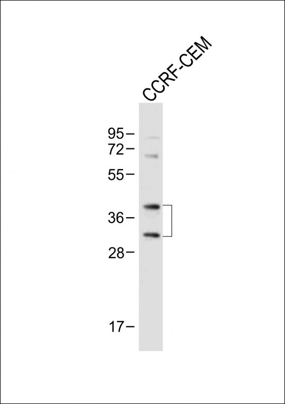 |
Anti-Mouse Nkx2-5 Antibody (Center) at 1:1000 dilution + CCRF-CEM whole cell lysate
Lysates/proteins at 20 µg per lane. Secondary Predicted band size : 30-42 kDa Blocking/Dilution buffer: 5% NFDM/TBST. |
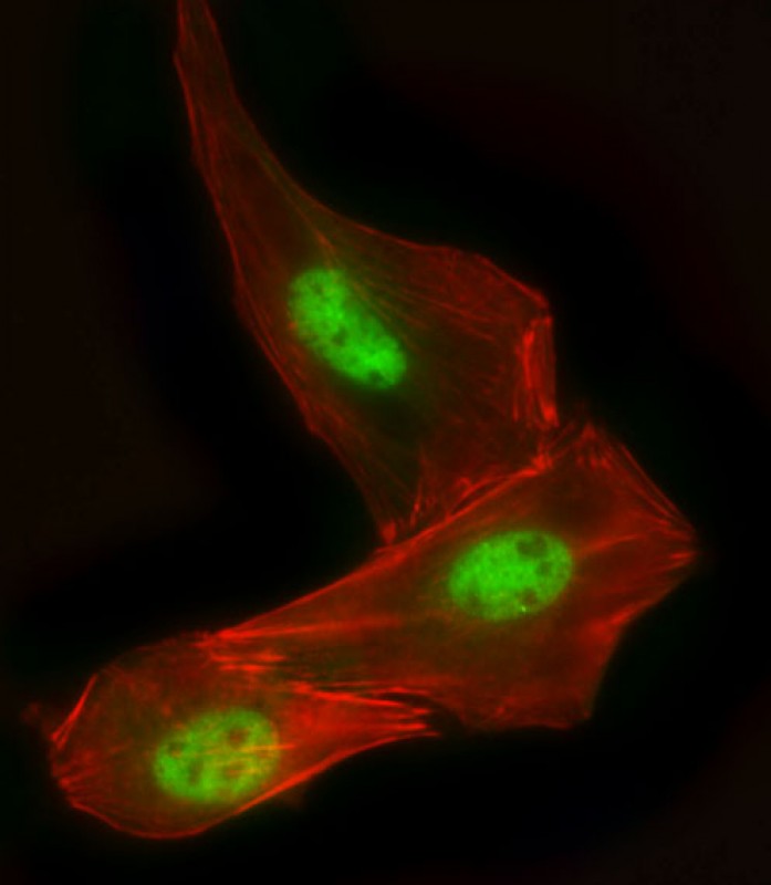 |
Immunofluorescent analysis of 4% paraformaldehyde-fixed, 0. 1% Triton X-100 permeabilized C2C12 cells labeling Nkx2-5 with P34700 at 1/25 dilution, followed by Dylight® 488-conjugated goat anti-Rabbit IgG secondary antibody at 1/200 dilution (green). Immunofluorescence image showing Nucleus staining on C2C12 cell line. Cytoplasmic actin is detected with Dylight® 554 Phalloidin(red). The nuclear counter stain is DAPI (blue). |
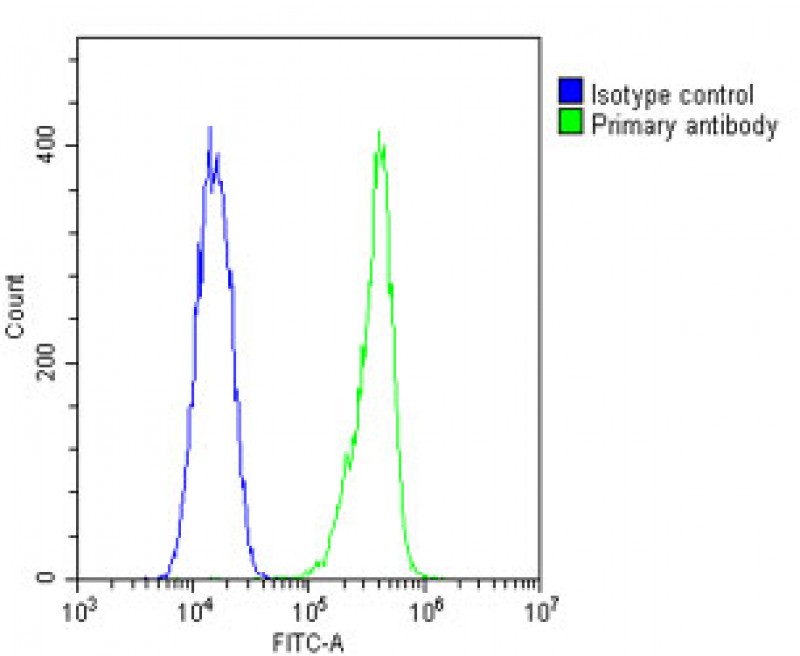 |
Overlay histogram showing C2C12 cells stained with P34700(green line). The cells were fixed with 2% paraformaldehyde and then permeabilized with 90% methanol for 10 min. The cells were then incubated in 2% bovine serum albumin to block non-specific protein-protein interactions followed by the antibody (1:25 dilution) for 60 min at 37ºC. The secondary antibody used was Goat-Anti-Rabbit IgG, DyLight® 488 Conjugated Highly Cross-Adsorbed at 1/200 dilution for 40 min at Room temperature. Isotype control antibody (blue line) was rabbit IgG1 (1μg/1×10^6 cells) used under the same conditions. Acquisition of >10, 000 events was performed. |
本公司的所有产品仅用于科学研究或者工业应用等非医疗目的,不可用于人类或动物的临床诊断或治疗,非药用,非食用。
暂无评论
本公司的所有产品仅用于科学研究或者工业应用等非医疗目的,不可用于人类或动物的临床诊断或治疗,非药用,非食用。


发表回复