 中文
中文 别名:应用:WB,IHC,ICC,FCM
反应种属:Human, Mouse, Rat
规格:50μl/100μl
| Specification | |
|---|---|
| Entrez GeneID | 20423 |
| Swissprot | Q62226 |
| WB Predicted band size | 47.8kDa |
| Host/Isotype | Rabbit IgG |
| Storage | Store at 4°C short term. Aliquot and store at -20°C long term. Avoid freeze/thaw cycles. |
| Species Reactivity | Human, Mouse, Rat |
| Immunogen | This mouse Shh antibody is generated from a rabbit immunized with a KLH conjugated synthetic peptide between 58-91 amino acids from the N-terminal region of mouse Shh. |
| Application | |
|---|---|
| WB | 1/2000 |
| IHC | 1/100-1/500 |
| ICC | 1/25 |
| FCM | 1/25 |
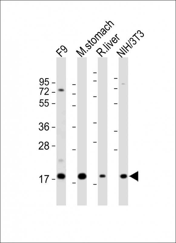 |
All lanes : Anti-Shh Antibody (N-term) at 1:2000 dilution Lane 1: F9 whole cell lysate Lane 2: mouse stomach lysates Lane 3: rat liver whole cell lysates Lane 4: NIH/3T3 lysates Lysates/proteins at 20 µg per lane. Secondary Goat Anti-Rabbit IgG, (H+L), Peroxidase conjugated at 1/10000 dilution Predicted band size : 48 kDa Blocking/Dilution buffer: 5% NFDM/TBST. Lysates/proteins at 20 µg per lane. Secondary Predicted band size : 84 kDa Blocking/Dilution buffer: 5% NFDM/TBST. |
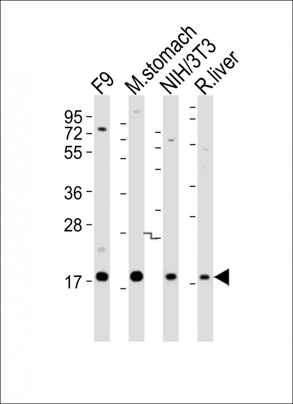 |
All lanes : Anti-Shh Antibody (N-term) at 1:2000 dilution Lane 1: F9 whole cell lysates Lane 2: mouse stomach lysates Lane 3: NIH/3T3 whole cell lysates Lane 4: rat liver lysates Lysates/proteins at 20 µg per lane. Secondary Predicted band size : 48 kDa Blocking/Dilution buffer: 5% NFDM/TBST. |
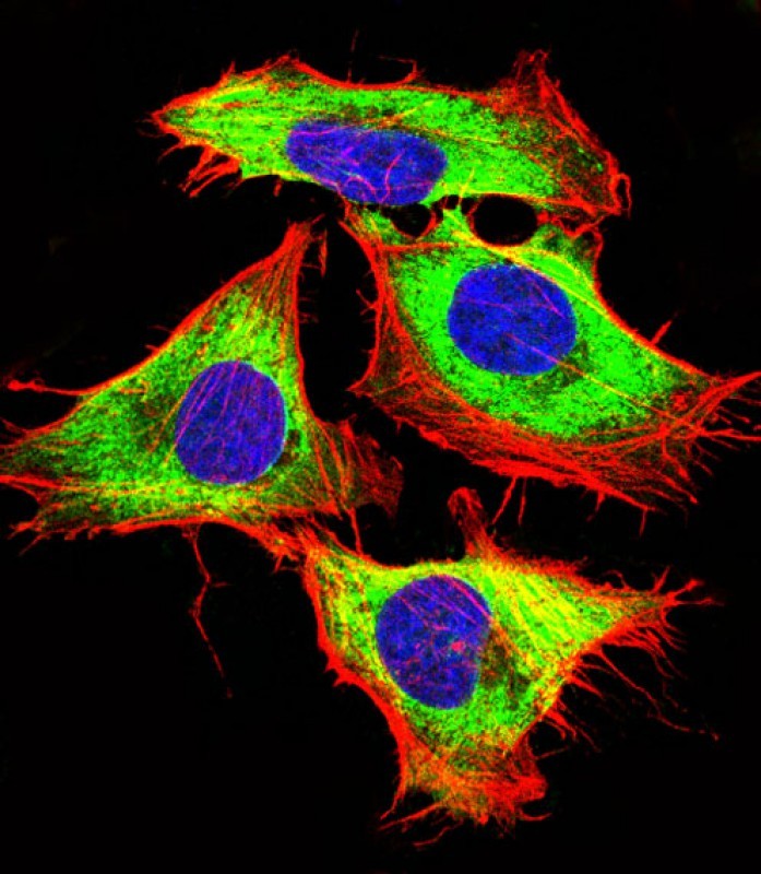 |
Immunofluorescent analysis of 4% paraformaldehyde-fixed, 0.1% Triton X-100 permeabilized Hela (Human Cervical epithelial adenocarcinoma cell line) cells labeling Shh with P34284 at 1/25 dilution, followed by Dylight® 488-conjugated goat anti-rabbit IgG secondary antibody at 1/200 dilution (green). Immunofluorescence image showing cytoplasm and membrane staining on Hela cell line. Cytoplasmic actin is detected with Dylight® 554 Phalloidin at 1/100 dilution (red).The nuclear counter stain is DAPI (blue). |
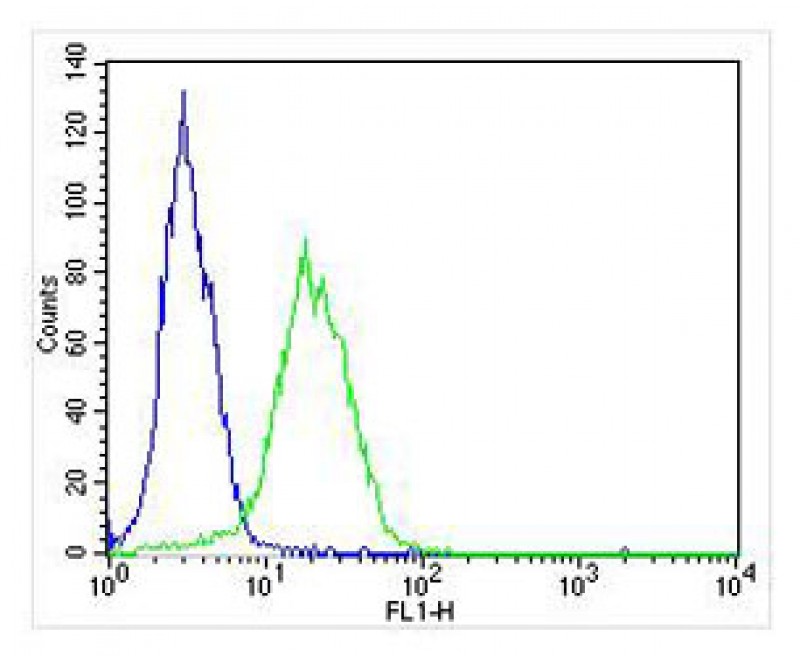 |
Overlay histogram showing HT-29 cells stained with P34284 (green line). The cells were fixed with 4% paraformaldehyde (10 min) and then permeabilized with 90% methanol for 10 min. The cells were then icubated in 2% bovine serum albumin to block non-specific protein-protein interactions followed by the antibody (AP12735b, 1:25 dilution) for 60 min at 37ºC. The secondary antibody used was Alexa Fluor® 488 goat anti-rabbit lgG (H+L) (1583138) at 1/400 dilution for 40 min at 37ºC. Isotype control antibody (blue line) was rabbit IgG1 (1μg/1×10^6 cells) used under the same conditions. Acquisition of >10, 000 events was performed. |
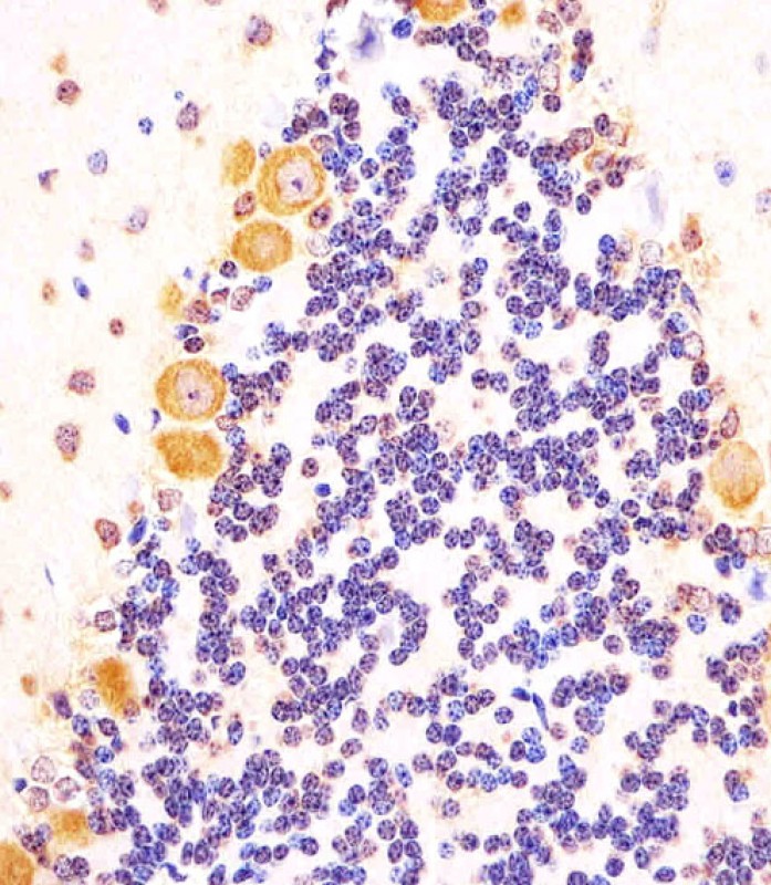 |
P34284 staining (Mouse) Shh in mouse cerebellum sections by Immunohistochemistry (IHC-P – paraformaldehyde-fixed, paraffin-embedded sections). Tissue was fixed with formaldehyde and blocked with 3% BSA for 0. 5 hour at room temperature; antigen retrieval was by heat mediation with a citrate buffer (pH6). Samples were incubated with primary antibody (1/25) for 1 hours at 37°C. A undiluted biotinylated goat polyvalent antibody was used as the secondary antibody. |
本公司的所有产品仅用于科学研究或者工业应用等非医疗目的,不可用于人类或动物的临床诊断或治疗,非药用,非食用。
暂无评论
本公司的所有产品仅用于科学研究或者工业应用等非医疗目的,不可用于人类或动物的临床诊断或治疗,非药用,非食用。


发表回复