 中文
中文 别名:Homeobox protein MSX-1, Homeobox protein Hox-7, Msh homeobox 1-like protein, MSX1, HOX7应用:WB,ICC
反应种属:Human, Mouse, Rat
规格:50μl/100μl
| Description |
|---|
| This gene encodes a member of the muscle segment homeobox gene family. The encoded protein functions as a transcriptional repressor during embryogenesis through interactions with components of the core transcription complex and other homeoproteins. It may also have roles in limb-pattern formation, craniofacial development, particularly odontogenesis, and tumor growth inhibition. Mutations in this gene, which was once known as homeobox 7, have been associated with nonsyndromic cleft lip with or without cleft palate 5, Witkop syndrome, Wolf-Hirschom syndrome, and autosomoal dominant hypodontia. |
| Specification | |
|---|---|
| Aliases | Homeobox protein MSX-1, Homeobox protein Hox-7, Msh homeobox 1-like protein, MSX1, HOX7 |
| Entrez GeneID | 4487 |
| Swissprot | P28360 |
| WB Predicted band size | 31.5kDa |
| Host/Isotype | Rabbit IgG |
| Storage | Store at 4°C short term. Aliquot and store at -20°C long term. Avoid freeze/thaw cycles. |
| Species Reactivity | Human, Mouse, Rat |
| Immunogen | This MSX1 antibody is generated from rabbits immunized with a KLH conjugated synthetic peptide between 111-138 amino acids from the Central region of human MSX1. |
| Formulation | Purified polyclonal antibody supplied in PBS with 0.05% sodium azide. This antibody is purified through a protein A column, followed by peptide affinity purification. |
| Application | |
|---|---|
| WB | 1/1000-1/2000 |
| ICC | 1/10-1/50 |
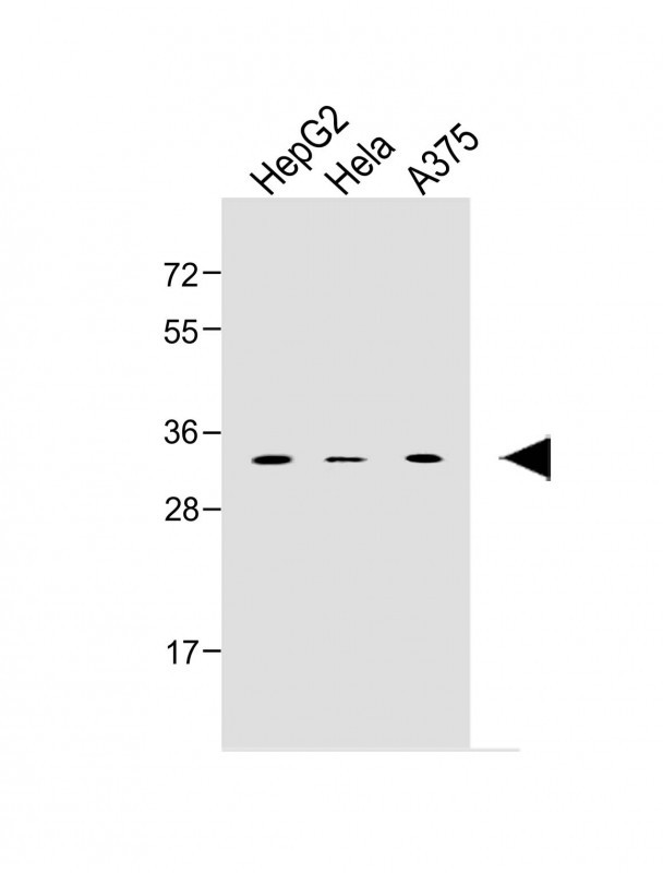 |
All lanes : Anti-MSX1 Antibody (Center) at 1:1000 dilution Lane 1: HepG2 MG whole cell lysate Lane 2: Hela whole cell lysate Lane 3: A375 whole cell lysate Lysates/proteins at 20 µg per lane. Secondary Observed band size : 32kDa Blocking/Dilution buffer: 5% NFDM/TBST. |
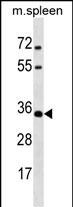 |
MSX1 Antibody (Center) (Cat. #P34893) western blot analysis in mouse spleen tissue lysates (35ug/lane).This demonstrates the MSX1 antibody detected the MSX1 protein (arrow). |
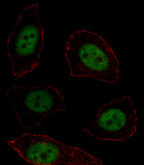 |
Fluorescent image of U251 cell stained with MSX1 Antibody (Center)(Cat#P34893).U251 cells were fixed with 4% PFA (20 min), permeabilized with Triton X-100 (0.1%, 10 min), then incubated with MSX1 primary antibody (1:25, 1 h at 37℃). For secondary antibody, Alexa Fluor® 488 conjugated donkey anti-rabbit antibody (green) was used (1:400, 50 min at 37℃).Cytoplasmic actin was counterstained with Alexa Fluor® 555 (red) conjugated Phalloidin (7units/ml, 1 h at 37℃).MSX1 immunoreactivity is localized to Nucleus significantly. |
本公司的所有产品仅用于科学研究或者工业应用等非医疗目的,不可用于人类或动物的临床诊断或治疗,非药用,非食用。
暂无评论
本公司的所有产品仅用于科学研究或者工业应用等非医疗目的,不可用于人类或动物的临床诊断或治疗,非药用,非食用。


发表回复