 中文
中文 别名:NADPH oxidase 1, NOX-1, 1—, Mitogenic oxidase 1, MOX-1, NADH/NADPH mitogenic oxidase subunit P65-MOX, NOH-1, NOX1, MOX1, NOH1应用:WB,IHC
反应种属:Human
规格:50μl/100μl
| Description |
|---|
| Voltage-gated proton (hydrogen) channels play an important role in cellular defense against acidic stress. They are unique among ion channels with respect to their extremely high selectivity, marked temperature dependence, and unitary conductance, which is 3 orders of magnitude lower than that of most other ion channels. NOX1 is a homolog of the catalytic subunit of the superoxide-generating NADPH oxidase of phagocytes, gp91phox. Two transcript variants encoding different isoforms have been found for this gene. |
| Specification | |
|---|---|
| Aliases | NADPH oxidase 1, NOX-1, 1—, Mitogenic oxidase 1, MOX-1, NADH/NADPH mitogenic oxidase subunit P65-MOX, NOH-1, NOX1, MOX1, NOH1 |
| Entrez GeneID | 27035 |
| Swissprot | Q9Y5S8 |
| WB Predicted band size | 64.9kDa |
| Host/Isotype | Rabbit IgG |
| Storage | Store at 4°C short term. Aliquot and store at -20°C long term. Avoid freeze/thaw cycles. |
| Species Reactivity | Human |
| Immunogen | This NOX1 antibody is generated from rabbits immunized with a KLH conjugated synthetic peptide between 243-271 amino acids from the Central region of human NOX1. |
| Formulation | Purified polyclonal antibody supplied in PBS with 0.05% sodium azide. This antibody is purified through a protein A column, followed by peptide affinity purification. |
| Application | |
|---|---|
| WB | 1/1000 |
| IHC | 1/500 |
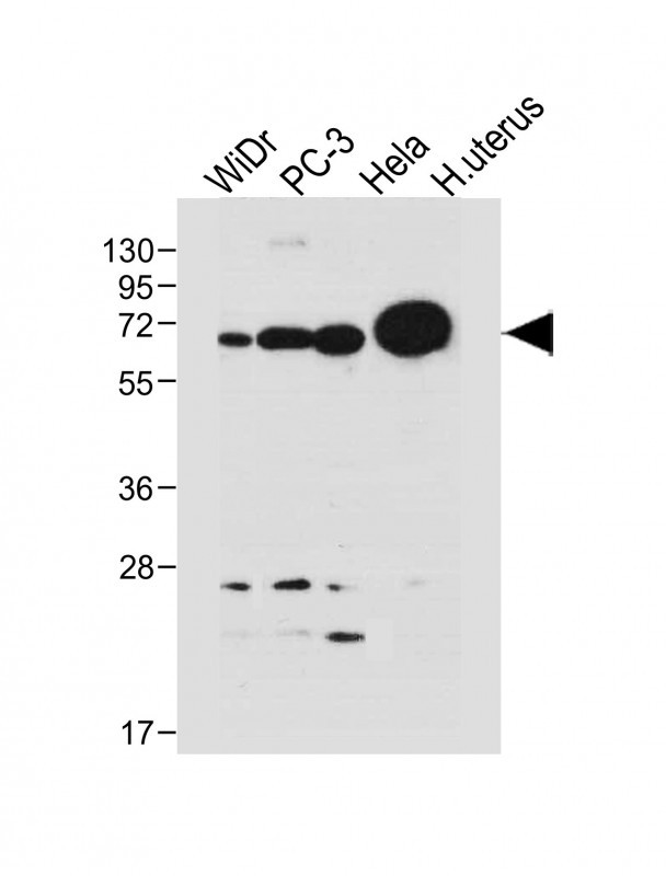 |
All lanes : Anti-NOX1 Antibody (Center) at 1:1000 dilution Lane 1: WiDr whole cell lysate Lane 2: PC-3 whole cell lysate Lane 3: Hela whole cell lysate Lane 4: human uterus tissue lysate Lysates/proteins at 20 µg per lane. Secondary Predicted band size : 65 kDa Blocking/Dilution buffer: 5% NFDM/TBST. |
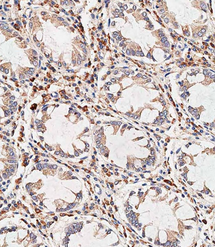 |
Immunohistochemical analysis of paraffin-embedded human colon tissue using P34662 performed on the Leica® BOND RXm. Tissue was fixed with formaldehyde at room temperature, antigen retrieval was by heat mediation with a EDTA buffer (pH9. 0). Samples were incubated with primary antibody(1:500) for 1 hours at room temperature. A undiluted biotinylated CRF Anti-Polyvalent HRP Polymer antibody was used as the secondary antibody. |
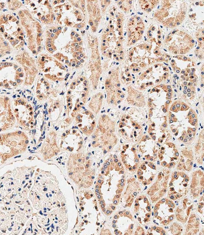 |
Immunohistochemical analysis of paraffin-embedded human kidney tissue using P34662 performed on the Leica® BOND RXm. Tissue was fixed with formaldehyde at room temperature, antigen retrieval was by heat mediation with a EDTA buffer (pH9. 0). Samples were incubated with primary antibody(1:500) for 1 hours at room temperature. A undiluted biotinylated CRF Anti-Polyvalent HRP Polymer antibody was used as the secondary antibody. |
本公司的所有产品仅用于科学研究或者工业应用等非医疗目的,不可用于人类或动物的临床诊断或治疗,非药用,非食用。
暂无评论
本公司的所有产品仅用于科学研究或者工业应用等非医疗目的,不可用于人类或动物的临床诊断或治疗,非药用,非食用。


发表回复