 中文
中文 别名:Niemann-Pick C1 protein, NPC1应用:WB,IHC,ICC
反应种属:Human, Mouse
规格:50μl/100μl
| Description |
|---|
| This gene encodes a large protein that resides in the limiting membrane of endosomes and lysosomes and mediates intracellular cholesterol trafficking via binding of cholesterol to its N-terminal domain. It is predicted to have a cytoplasmic C-terminus, 13 transmembrane domains, and 3 large loops in the lumen of the endosome – the last loop being at the N-terminus. This protein transports low-density lipoproteins to late endosomal/lysosomal compartments where they are hydrolized and released as free cholesterol. Defects in this gene cause Niemann-Pick type C disease, a rare autosomal recessive neurodegenerative disorder characterized by over accumulation of cholesterol and glycosphingolipids in late endosomal/lysosomal compartments. |
| Specification | |
|---|---|
| Aliases | Niemann-Pick C1 protein, NPC1 |
| Entrez GeneID | 4864 |
| Swissprot | O15118 |
| WB Predicted band size | 142.2kDa |
| Host/Isotype | Rabbit IgG |
| Storage | Store at 4°C short term. Aliquot and store at -20°C long term. Avoid freeze/thaw cycles. |
| Species Reactivity | Human, Mouse |
| Immunogen | This NPC1 antibody is generated from rabbits immunized with a KLH conjugated synthetic peptide between 591-620 amino acids from the Central region of human NPC1. |
| Formulation | Purified polyclonal antibody supplied in PBS with 0.05% sodium azide. This antibody is purified through a protein A column, followed by peptide affinity purification. |
| Application | |
|---|---|
| WB | 1/1000-1/2000 |
| IHC | 1/100-1/500 |
| ICC | 1/10-1/50 |
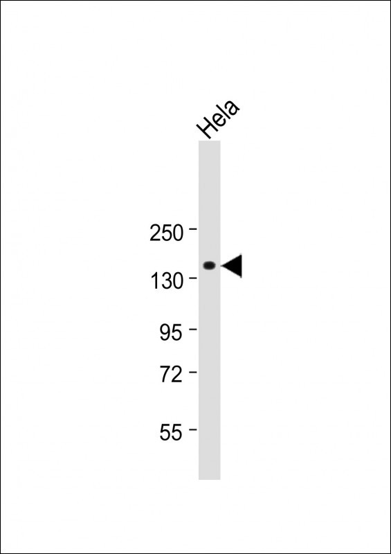 |
Anti-NPC1 Antibody (Center) at 1:2000 dilution + Hela whole cell lysate
Lysates/proteins at 20 µg per lane. Secondary Predicted band size : 142 kDa Blocking/Dilution buffer: 5% NFDM/TBST. |
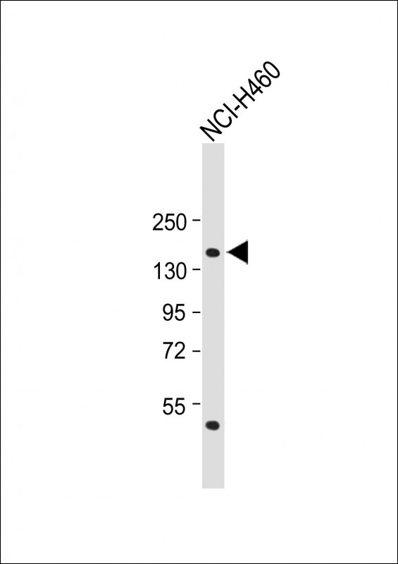 |
Anti-NPC1 Antibody (Center) at 1:1000 dilution + NCI-H460 whole cell lysate
Lysates/proteins at 20 µg per lane. Secondary Predicted band size : 142 kDa Blocking/Dilution buffer: 5% NFDM/TBST. |
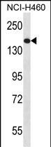 |
NPC1 Antibody (Center) (Cat. #P34840) western blot analysis in NCI-H460 cell line lysates (35ug/lane).This demonstrates the NPC1 antibody detected the NPC1 protein (arrow). |
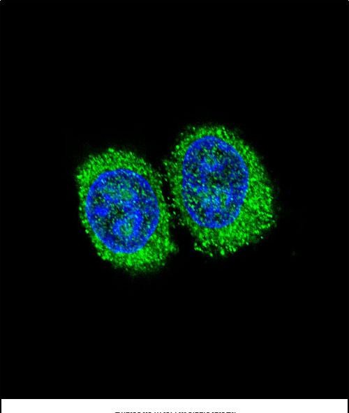 |
Confocal immunofluorescent analysis of NPC1 Antibody (Center) (Cat#P34840) with 293 cell followed by Alexa Fluor 488-conjugated goat anti-rabbit lgG (green). DAPI was used to stain the cell nuclear (blue). |
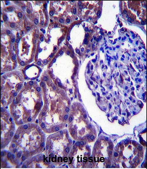 |
NPC1 Antibody (Center) (Cat. #P34840)immunohistochemistry analysis in formalin fixed and paraffin embedded human kidney tissue followed by peroxidase conjugation of the secondary antibody and DAB staining.This data demonstrates the use of NPC1 Antibody (Center) for immunohistochemistry. Clinical relevance has not been evaluated. |
本公司的所有产品仅用于科学研究或者工业应用等非医疗目的,不可用于人类或动物的临床诊断或治疗,非药用,非食用。
暂无评论
本公司的所有产品仅用于科学研究或者工业应用等非医疗目的,不可用于人类或动物的临床诊断或治疗,非药用,非食用。


发表回复