 中文
中文 别名:Protein disulfide-isomerase A6, Endoplasmic reticulum protein 5, ER protein 5, ERp5, Protein disulfide isomerase P5, Thioredoxin domain-containing protein 7, PDIA6, ERP5, P5, TXNDC7应用:WB,IHC,ICC
反应种属:Human, Mouse, Rat
规格:50μl/100μl
| Description |
|---|
| Protein disulfide isomerases (EC 5.3.4.1), such as PDIA6, are endoplasmic reticulum (ER) resident proteins that catalyze formation, reduction, and isomerization of disulfide bonds in proteins and are thought to play a role in folding of disulfide-bonded proteins. |
| Specification | |
|---|---|
| Aliases | Protein disulfide-isomerase A6, Endoplasmic reticulum protein 5, ER protein 5, ERp5, Protein disulfide isomerase P5, Thioredoxin domain-containing protein 7, PDIA6, ERP5, P5, TXNDC7 |
| Entrez GeneID | 10130 |
| Swissprot | Q15084 |
| WB Predicted band size | 48.1kDa |
| Host/Isotype | Rabbit IgG |
| Storage | Store at 4°C short term. Aliquot and store at -20°C long term. Avoid freeze/thaw cycles. |
| Species Reactivity | Human, Mouse, Rat |
| Immunogen | This PDIA6 antibody is generated from rabbits immunized with a KLH conjugated synthetic peptide between 144-172 amino acids from the Central region of human PDIA6. |
| Formulation | Purified polyclonal antibody supplied in PBS with 0.05% sodium azide. This antibody is prepared by Saturated Ammonium Sulfate (SAS) precipitation followed by dialysis against PBS. |
| Application | |
|---|---|
| WB | 1/1000 |
| IHC | 1/100-1/500 |
| ICC | 1/100 |
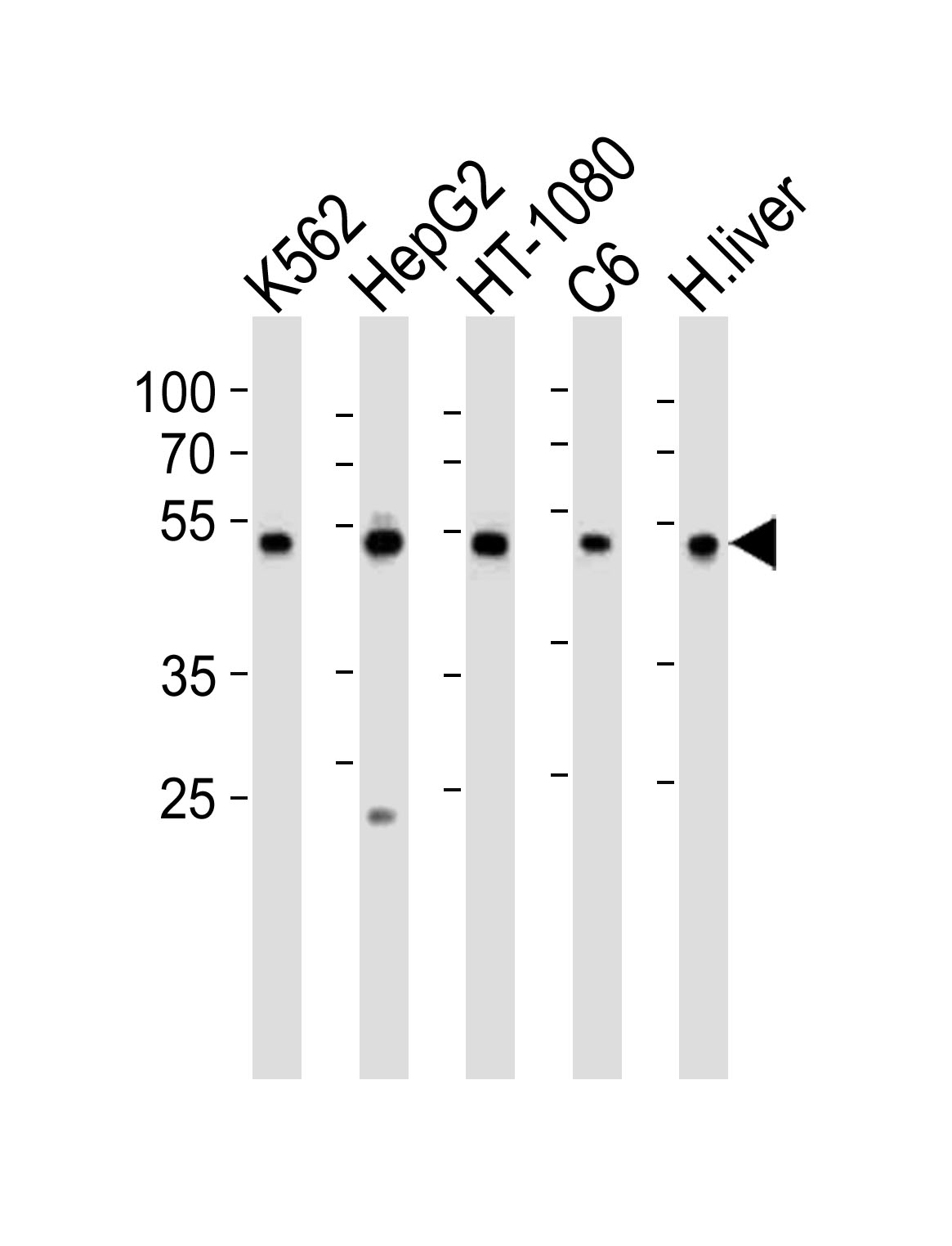 |
Western blot analysis of lysates from K562, HepG2, HT-1080, rat C6 cell line and human liver tissue lysate (from left to right), using PDIA6 Antibody (Center K159)(Cat. #P32745). P32745 was diluted at 1:1000 at each lane. A goat anti-rabbit IgG H&L(HRP) at 1:10000 dilution was used as the secondary antibody. Lysates at 35ug per lane. |
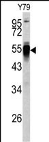 |
Western blot analysis of PDIA6 antibody (Center K159) (Cat.# P32745) in Y79 cell line lysates (35ug/lane). PDIA6 (arrow) was detected using the purified Pab. |
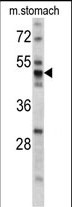 |
Western blot analysis of PDIA6 antibody (Center K159) (Cat.# P32745) in mouse stomach tissue lysates (35ug/lane). PDIA6 (arrow) was detected using the purified Pab. |
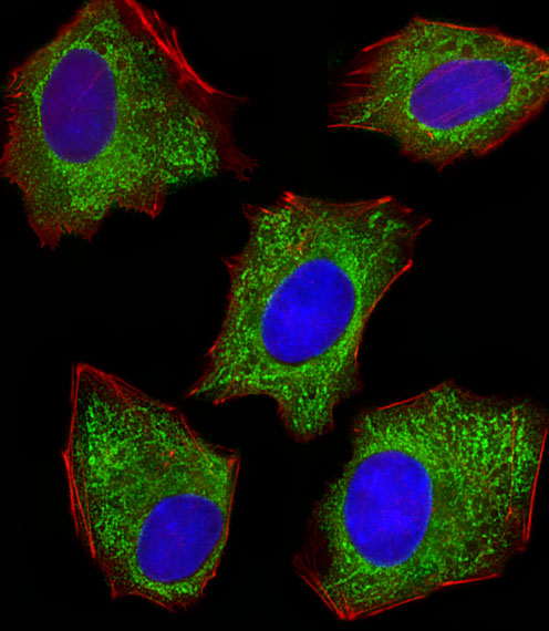 |
Fluorescent image of HepG2 cells stained with XAF1 PDIA6 Antibody (Center K159)(Cat#P32745). P32745 was diluted at 1:100 dilution. An Alexa Fluor 488-conjugated goat anti-rabbit lgG at 1:400 dilution was used as the secondary antibody (green). DAPI was used to stain the cell nuclear (blue). Cytoplasmic actin was counterstained with Alexa Fluor® 555 conjugated with Phalloidin (red). |
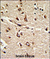 |
Formalin-fixed and paraffin-embedded human brain tissue reacted with PDIA6 Antibody (Center K159), which was peroxidase-conjugated to the secondary antibody, followed by DAB staining. This data demonstrates the use of this antibody for immunohistochemistry; clinical relevance has not been evaluated. |
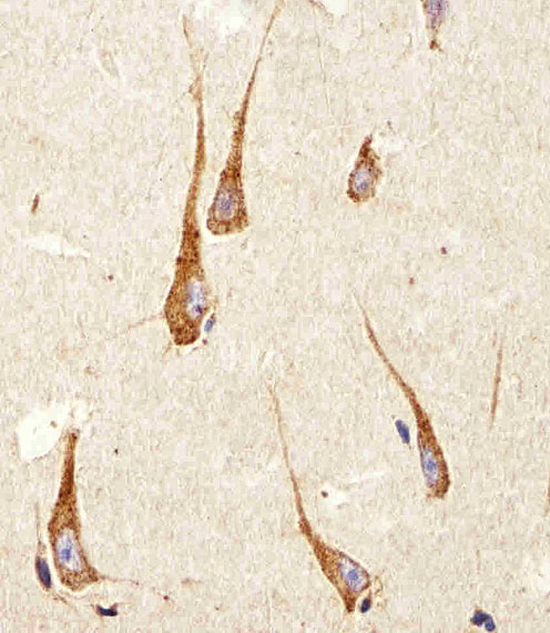 |
Immunohistochemical analysis of paraffin-embedded H. brain section using PDIA6 Antibody (Center K159)(Cat#P32745). P32745 was diluted at 1:100 dilution. A peroxidase-conjugated goat anti-rabbit IgG at 1:400 dilution was used as the secondary antibody, followed by DAB staining. |
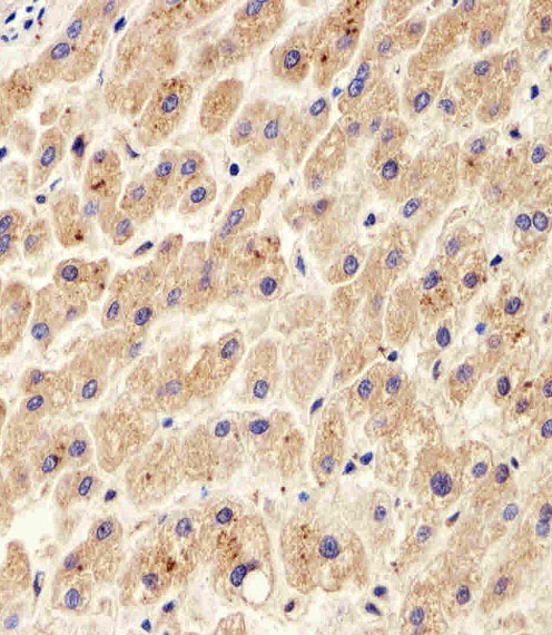 |
Immunohistochemical analysis of paraffin-embedded H. liver section using PDIA6 Antibody (Center K159)(Cat#P32745). P32745 was diluted at 1:100 dilution. A peroxidase-conjugated goat anti-rabbit IgG at 1:400 dilution was used as the secondary antibody, followed by DAB staining. |
本公司的所有产品仅用于科学研究或者工业应用等非医疗目的,不可用于人类或动物的临床诊断或治疗,非药用,非食用。
暂无评论
本公司的所有产品仅用于科学研究或者工业应用等非医疗目的,不可用于人类或动物的临床诊断或治疗,非药用,非食用。


发表回复