 中文
中文 别名:GPI inositol-deacylase, 3.1.-.-, Post-GPI attachment to proteins factor 1, hPGAP1, PGAP1应用:WB,ICC,FCM
反应种属:Human, Mouse, Rat
规格:50μl/100μl
| Description |
|---|
| Involved in inositol deacylation of GPI-anchored proteins. GPI inositol deacylation may important for efficient transport of GPI-anchored proteins from the endoplasmic reticulum to the Golgi (By similarity). |
| Specification | |
|---|---|
| Aliases | GPI inositol-deacylase, 3.1.-.-, Post-GPI attachment to proteins factor 1, hPGAP1, PGAP1 |
| Entrez GeneID | 80055 |
| Swissprot | Q75T13 |
| WB Predicted band size | 105.4kDa |
| Host/Isotype | Rabbit IgG |
| Storage | Store at 4°C short term. Aliquot and store at -20°C long term. Avoid freeze/thaw cycles. |
| Species Reactivity | Human, Mouse, Rat |
| Immunogen | This PGAP1 antibody is generated from a rabbit immunized with a KLH conjugated synthetic peptide between 90-122 amino acids from human PGAP1. |
| Application | |
|---|---|
| WB | 1/2000 |
| ICC | 1/25 |
| FCM | 1/25 |
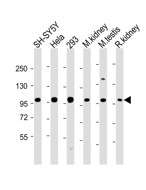 |
All lanes : Anti-PGAP1 Antibody (N-Term) at 1:2000 dilution Lane 1: SH-SY5Y whole cell lysate Lane 2: Hela whole cell lysate Lane 3: 293 whole cell lysate Lane 4: mouse kidney lysate Lane 5: mouse testis lysate Lane 6: rat kidney lysate Lysates/proteins at 20 µg per lane. Secondary Predicted band size : 105 kDa Blocking/Dilution buffer: 5% NFDM/TBST. |
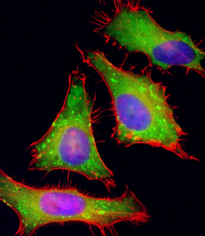 |
Immunofluorescent analysis of 4% paraformaldehyde-fixed, 0.1% Triton X-100 permeabilized HeLa (human cervical epithelial adenocarcinoma cell line) cells labeling PGAP1 with P34362 at 1/25 dilution, followed by Dylight® 488-conjugated goat anti-rabbit IgG secondary antibody at 1/200 dilution (green). Immunofluorescence image showing cytoplasm staining on HeLa cell line. Cytoplasmic actin is detected with Dylight® 554 Phalloidin at 1/100 dilution (red).The nuclear counter stain is DAPI (blue). |
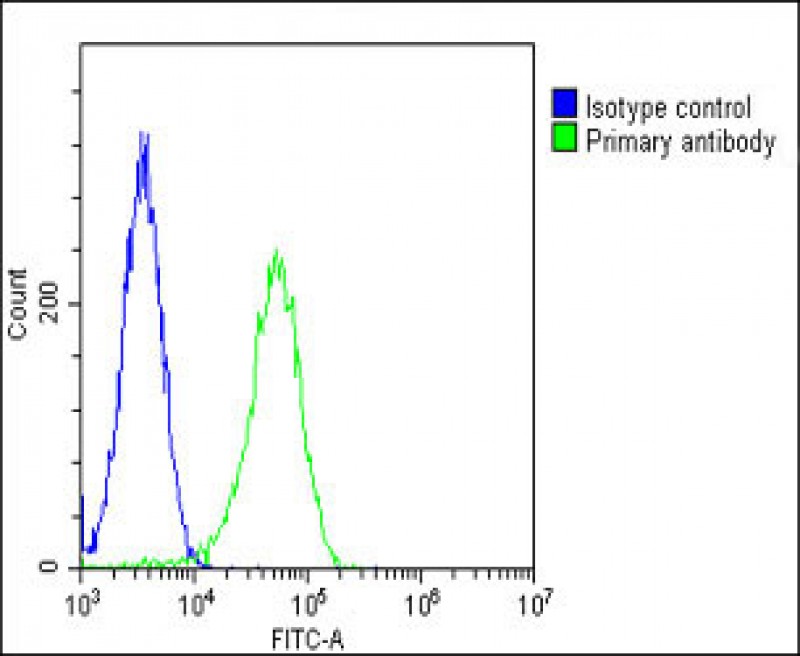 |
Overlay histogram showing Hela cells stained with P34362(green line). The cells were fixed with 2% paraformaldehyde (10 min) and then permeabilized with 90% methanol for 10 min. The cells were then icubated in 2% bovine serum albumin to block non-specific protein-protein interactions followed by the antibody (P34362, 1:25 dilution) for 60 min at 37ºC. The secondary antibody used was Goat-Anti-Rabbit IgG, DyLight®488 Conjugated Highly Cross-Adsorbed(OH191631) at 1/200 dilution for 40 min at 37ºC. Isotype control antibody (blue line) was rabbit IgG1 (1μg/1×10^6 cells) used under the same conditions. Acquisition of >10, 000 events was performed. |
本公司的所有产品仅用于科学研究或者工业应用等非医疗目的,不可用于人类或动物的临床诊断或治疗,非药用,非食用。
暂无评论
本公司的所有产品仅用于科学研究或者工业应用等非医疗目的,不可用于人类或动物的临床诊断或治疗,非药用,非食用。


发表回复