 中文
中文 别名:M-phase inducer phosphatase 1, Dual specificity phosphatase Cdc25A, CDC25A应用:WB,IHC,ICC
反应种属:Human, Mouse, Rat
规格:50μl/100μl
| Description |
|---|
| CDC25A is a member of the CDC25 family of phosphatases. CDC25A is required for progression from G1 to the S phase of the cell cycle. It activates the cyclin-dependent kinase CDC2 by removing two phosphate groups. CDC25A is specifically degraded in response to DNA damage, which prevents cells with chromosomal abnormalities from progressing through cell division. CDC25A is an oncogene, although its exact role in oncogenesis has not been demonstrated. Two transcript variants encoding different isoforms have been found for this gene. |
| Specification | |
|---|---|
| Aliases | M-phase inducer phosphatase 1, Dual specificity phosphatase Cdc25A, CDC25A |
| Entrez GeneID | 993 |
| Swissprot | P30304 |
| WB Predicted band size | 59.1kDa |
| Host/Isotype | Rabbit IgG |
| Storage | Store at 4°C short term. Aliquot and store at -20°C long term. Avoid freeze/thaw cycles. |
| Species Reactivity | Human, Mouse, Rat |
| Immunogen | This CDC25A Antibody is generated from rabbits immunized with a KLH conjugated synthetic phosphopeptide corresponding to amino acid residues surrounding T507 of human CDC25A. |
| Formulation | Purified polyclonal antibody supplied in PBS with 0.05% sodium azide. This antibody is purified through a protein A column, followed by peptide affinity purification. |
| Application | |
|---|---|
| WB | 1/1000 |
| IHC | 1/100-1/500 |
| ICC | 1/25 |
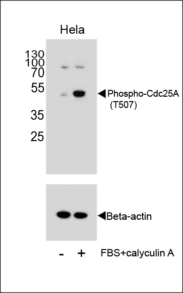 |
Western blot analysis of extracts from Hela cells, untreated or treated with calyculin A, using Phospho-Cdc25A Antibody (T507). |
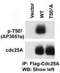 |
The anti-Phospho-CDC25A-T507 Pab (Cat. #P32633) is used in Western blot to detect Phospho-CDC25A-T507 in cells transfected with wild type or mutant T507 A of CDC25A. Data courtesy of Dr. Tiebang Kang of Washington University, St. Louis, MO. |
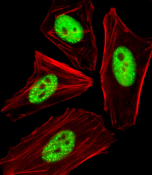 |
Fluorescent image of HeLa cells stained with Phospho-CDC25A(T507) Antibody(Cat#P32633). P32633 was diluted at 1:25 dilution. An Alexa Fluor 488-conjugated goat anti-rabbit lgG at 1:400 dilution was used as the secondary antibody (green). Cytoplasmic actin was counterstained with Alexa Fluor® 555 conjugated with Phalloidin (red). |
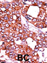 |
Formalin-fixed and paraffin-embedded human cancer tissue reacted with the primary antibody, which was peroxidase-conjugated to the secondary antibody, followed by AEC staining. This data demonstrates the use of this antibody for immunohistochemistry; clinical relevance has not been evaluated. BC = breast carcinoma; HC = hepatocarcinoma. |
本公司的所有产品仅用于科学研究或者工业应用等非医疗目的,不可用于人类或动物的临床诊断或治疗,非药用,非食用。
暂无评论
本公司的所有产品仅用于科学研究或者工业应用等非医疗目的,不可用于人类或动物的临床诊断或治疗,非药用,非食用。


发表回复