 中文
中文 别名:Plectin, PCN, PLTN, Hemidesmosomal protein 1, HD1, Plectin-1, PLEC, PLEC1应用:WB,ICC
反应种属:Human, Mouse, Rat
规格:50μl/100μl
| Description |
|---|
| Interlinks intermediate filaments with microtubules and microfilaments and anchors intermediate filaments to desmosomes or hemidesmosomes. Could also bind muscle proteins such as actin to membrane complexes in muscle. May be involved not only in the filaments network, but also in the regulation of their dynamics. Structural component of muscle. Isoform 9 plays a major role in the maintenance of myofibers integrity. |
| Specification | |
|---|---|
| Aliases | Plectin, PCN, PLTN, Hemidesmosomal protein 1, HD1, Plectin-1, PLEC, PLEC1 |
| Entrez GeneID | 5339 |
| Swissprot | Q15149 |
| WB Predicted band size | 531.8kDa |
| Host/Isotype | Rabbit IgG |
| Storage | Store at 4°C short term. Aliquot and store at -20°C long term. Avoid freeze/thaw cycles. |
| Species Reactivity | Human, Mouse, Rat |
| Immunogen | This PLEC antibody is generated from a rabbit immunized with a KLH conjugated synthetic peptide between 4241-4275 amino acids from human PLEC. |
| Application | |
|---|---|
| WB | 1/1000 |
| ICC | 1/25 |
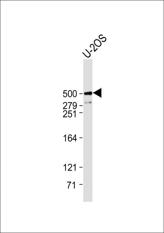 |
Anti-PLEC Antibody (C-Term) at 1:1000 dilution + U-2OS whole cell lysate
Lysates/proteins at 20 µg per lane. Secondary Predicted band size : 532 kDa Blocking/Dilution buffer: 5% NFDM/TBST. |
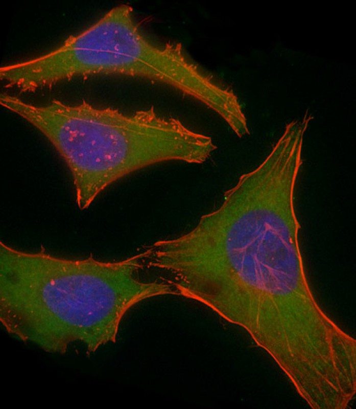 |
Immunofluorescent analysis of 4% paraformaldehyde-fixed, 0.1% Triton X-100 permeabilized HeLa (human cervical epithelial adenocarcinoma cell line) cells labeling PLEC with P34436 at 1/25 dilution, followed by Dylight® 488-conjugated goat anti-rabbit IgG secondary antibody at 1/200 dilution (green). Immunofluorescence image showing cytoplasm staining on HeLa cell line. Cytoplasmic actin is detected with Dylight® 554 Phalloidin at 1/100 dilution (red). The nuclear counter stain is DAPI (blue). |
本公司的所有产品仅用于科学研究或者工业应用等非医疗目的,不可用于人类或动物的临床诊断或治疗,非药用,非食用。
暂无评论
本公司的所有产品仅用于科学研究或者工业应用等非医疗目的,不可用于人类或动物的临床诊断或治疗,非药用,非食用。


发表回复