 中文
中文 别名:DNA polymerase alpha catalytic subunit, 2.7.7.7, DNA polymerase alpha catalytic subunit p180, POLA1, POLA应用:WB,FCM
反应种属:Human, Mouse
规格:50μl/100μl
| Description |
|---|
| Plays an essential role in the initiation of DNA replication. During the S phase of the cell cycle, the DNA polymerase alpha complex (composed of a catalytic subunit POLA1/p180, a regulatory subunit POLA2/p70 and two primase subunits PRIM1/p49 and PRIM2/p58) is recruited to DNA at the replicative forks via direct interactions with MCM10 and WDHD1. The primase subunit of the polymerase alpha complex initiates DNA synthesis by oligomerising short RNA primers on both leading and lagging strands. These primers are initially extended by the polymerase alpha catalytic subunit and subsequently transferred to polymerase delta and polymerase epsilon for processive synthesis on the lagging and leading strand, respectively. The reason this transfer occurs is because the polymerase alpha has limited processivity and lacks intrinsic 3′ exonuclease activity for proofreading error, and therefore is not well suited for replicating long complexes. |
| Specification | |
|---|---|
| Aliases | DNA polymerase alpha catalytic subunit, 2.7.7.7, DNA polymerase alpha catalytic subunit p180, POLA1, POLA |
| Entrez GeneID | 5422 |
| Swissprot | P09884 |
| WB Predicted band size | 165.9kDa |
| Host/Isotype | Rabbit IgG |
| Storage | Store at 4°C short term. Aliquot and store at -20°C long term. Avoid freeze/thaw cycles. |
| Species Reactivity | Human, Mouse |
| Immunogen | This POLA1 antibody is generated from a rabbit immunized with a KLH conjugated synthetic peptide between 1406-1439 amino acids from the human region of human POLA1. |
| Application | |
|---|---|
| WB | 1/2000 |
| FCM | 1/25 |
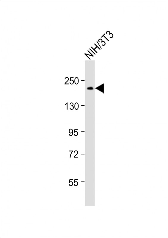 |
Anti-POLA1 Antibody (C-Term) at 1:2000 dilution + NIH/3T3 whole cell lysate
Lysates/proteins at 20 µg per lane. Secondary Predicted band size : 166 kDa Blocking/Dilution buffer: 5% NFDM/TBST. |
 |
Overlay histogram showing A431 cells stained with P34506(green line). The cells were fixed with 2% paraformaldehyde and then permeabilized with 90% methanol for 10 min. The cells were then icubated in 2% bovine serum albumin to block non-specific protein-protein interactions followed by the antibody (1:25 dilution) for 60 min at 37ºC. The secondary antibody used was Goat-Anti-Rabbit IgG, DyLight® 488 Conjugated Highly Cross-Adsorbed at 1/200 dilution for 40 min at Room temperature. Isotype control antibody (blue line) was rabbit IgG1 (1μg/1×10^6 cells) used under the same conditions. Acquisition of >10, 000 events was performed. |
本公司的所有产品仅用于科学研究或者工业应用等非医疗目的,不可用于人类或动物的临床诊断或治疗,非药用,非食用。
暂无评论
本公司的所有产品仅用于科学研究或者工业应用等非医疗目的,不可用于人类或动物的临床诊断或治疗,非药用,非食用。


发表回复