 中文
中文 别名:Retinoblastoma-like protein 2, 130 kDa retinoblastoma-associated protein, p130, Retinoblastoma-related protein 2, RBR-2, pRb2, RBL2, RB2应用:WB,FCM
反应种属:Human, Mouse, Rat
规格:50μl/100μl
| Description |
|---|
| RBL2 is a key regulator of entry into cell division. Directly involved in heterochromatin formation by maintaining overall chromatin structure and, in particular, that of constitutive heterochromatin by stabilizing histone methylation. Recruits and targets histone methyltransferases SUV420H1 and SUV420H2, leading to epigenetic transcriptional repression. Controls histone H4 ‘Lys-20’ trimethylation. Probably acts as a transcription repressor by recruiting chromatin-modifying enzymes to promoters. Potent inhibitor of E2F-mediated trans-activation, associates preferentially with E2F5. Binds to cyclins A and E. Binds to and may be involved in the transforming capacity of the adenovirus E1A protein. RBL2 may act as a tumor suppressor. |
| Specification | |
|---|---|
| Aliases | Retinoblastoma-like protein 2, 130 kDa retinoblastoma-associated protein, p130, Retinoblastoma-related protein 2, RBR-2, pRb2, RBL2, RB2 |
| Entrez GeneID | 5934 |
| Swissprot | Q08999 |
| WB Predicted band size | 128.4kDa |
| Host/Isotype | Rabbit IgG |
| Storage | Store at 4°C short term. Aliquot and store at -20°C long term. Avoid freeze/thaw cycles. |
| Species Reactivity | Human, Mouse, Rat |
| Immunogen | This RBL2 antibody is generated from rabbits immunized with a KLH conjugated synthetic peptide between 165-194 amino acids from the N-terminal region of human RBL2. |
| Formulation | Purified polyclonal antibody supplied in PBS with 0.05% sodium azide. This antibody is purified through a protein A column, followed by peptide affinity purification. |
| Application | |
|---|---|
| WB | 1/1000 |
| FCM | 1/10-1/50 |
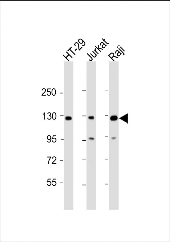 |
All lanes : Anti-RBL2 Antibody (N-term) at 1:2000 dilution Lane 1: HT-29 whole cell lysate Lane 2: Jurkat whole cell lysate Lane 3: Raji whole cell lysate Lysates/proteins at 20 µg per lane. Secondary Predicted band size : 128 kDa Blocking/Dilution buffer: 5% NFDM/TBST. |
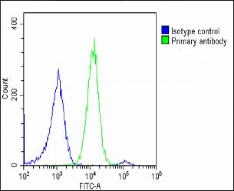 |
Overlay histogram showing U-2 OS cells stained with P34312(green line). The cells were fixed with 2% paraformaldehyde (10 min) and then permeabilized with 90% methanol for 10 min. The cells were then icubated in 2% bovine serum albumin to block non-specific protein-protein interactions followed by the antibody (P34312, 1:25 dilution) for 60 min at 37ºC. The secondary antibody used was Goat-Anti-Rabbit IgG, DyLight® 488 Conjugated Highly Cross-Adsorbed(OE188374) at 1/200 dilution for 40 min at 37ºC. Isotype control antibody (blue line) was rabbit IgG1 (1μg/1×10^6 cells) used under the same conditions. Acquisition of >10, 000 events was performed. |
本公司的所有产品仅用于科学研究或者工业应用等非医疗目的,不可用于人类或动物的临床诊断或治疗,非药用,非食用。
暂无评论
本公司的所有产品仅用于科学研究或者工业应用等非医疗目的,不可用于人类或动物的临床诊断或治疗,非药用,非食用。


发表回复